

Capnography in cardiac arrest turns a chaotic resuscitation into something you can measure. In the middle of a code, most feedback is delayed or subjective. The capnograph is neither.
It gives the team a live, physiological window into what the compressions are actually achieving. It confirms the airway, it grades the quality of CPR, and it announces the return of a pulse before your fingers can find one. This guide covers all three uses, and the one place where the numbers must be read with great care.
Key takeaways
- Capnography does three jobs in an arrest. It confirms the airway, measures CPR quality, and signals ROSC.
- EtCO₂ reflects the blood flow your compressions are generating. Higher is better.
- A value under 10 mmHg means compressions need to improve, not that you stop.
- A sudden sustained rise is often the first sign of ROSC, before a pulse is felt.
- For prognosis, a low value after 20 minutes is one factor among many, never used alone.
Why capnography belongs in every arrest
During cardiac arrest, the heart is not moving blood. The only circulation is the circulation your chest compressions create.
EtCO₂ measures the carbon dioxide returning to the lungs. That return depends on blood flow. So the number on the capnograph is a live estimate of the perfusion your compressions are generating. No other tool at the bedside gives you that, continuously, without stopping CPR.
The 2025 to 2030 American Heart Association ACLS guidelines recommend quantitative waveform capnography in intubated patients during cardiac arrest, for exactly these reasons.
Confirming the airway during CPR
The first job is the tube. Continuous waveform capnography, with clinical assessment, is the most reliable way to confirm and monitor correct endotracheal tube placement.
In a code this matters twice over. You confirm the tube at intubation, and you keep confirming it while the patient is moved, turned and transferred. A tube that slips out during a resuscitation is caught by the waveform at once. For the full method, see endotracheal tube confirmation.
One caution belongs here. In very low blood flow, a correctly placed tube can still show a low reading, because there is little perfusion to carry CO₂ to the lungs. Read the number in the context of the arrest.
Capnography as a measure of CPR quality
This is where capnography changes how a team performs.
Because EtCO₂ tracks the blood flow your compressions produce, it tells you whether those compressions are working. High-quality CPR generally produces an EtCO₂ of at least 10 to 20 mmHg. A value under 10 mmHg is a signal that the compressions need to improve, through greater depth, a better rate, full chest recoil or a fresh compressor.
A gradual fall in EtCO₂ over a few minutes is a classic sign of a tiring compressor. It is your cue to rotate. The number turns CPR quality from a guess into a target the whole team can see. For what the values mean in health, see the normal EtCO₂ range.
Detecting the return of spontaneous circulation
The most striking use is ROSC detection.
When the heart restarts, blood flow surges, more CO₂ reaches the lungs, and EtCO₂ jumps. A sudden sustained rise, often to near-normal values, is frequently the earliest sign of ROSC, appearing before a carotid pulse can be felt. If you see that spike during compressions, pause and check the pulse.
This has a real advantage. It lets the team recognise ROSC without repeatedly stopping compressions for pulse checks, which keeps perfusion going when it matters. After ROSC, EtCO₂ then helps you titrate ventilation to a normal target rather than over-ventilating a fragile patient.
Capnography and ROSC prediction
Here is the part that demands care. EtCO₂ during CPR carries prognostic information, and it is easy to misuse.
The evidence is consistent. A persistently low EtCO₂, under 10 mmHg after 20 minutes of high-quality CPR in an intubated patient, is associated with a very low likelihood of return of circulation, around half of one percent in several studies. Higher values, above 20 mmHg at 20 minutes, correlate with better outcomes and have even been proposed as a threshold to consider extracorporeal CPR.
But a number this powerful is also dangerous if read alone. The American Heart Association is explicit. A low EtCO₂ after 20 minutes may be considered as one part of a multimodal decision about when to stop, and it must never be used in isolation.
The reason is confounders. A low reading can also come from poor compressions, a dislodged or obstructed tube, a disconnected circuit, a pulmonary embolism, or the type of arrest itself. Each of these lowers EtCO₂ without meaning the patient is beyond help. So the value informs the decision. It never makes it.
What the guidelines say
The position is settled and consistent across editions.
Continuous waveform capnography is recommended to confirm and monitor tube placement, to monitor the quality of CPR and to help detect ROSC. In intubated patients, a failure to reach an EtCO₂ above 10 mmHg after 20 minutes of CPR may form one component of a multimodal decision to end efforts, but not the sole criterion. That single sentence captures both the value and the limit.
How to use capnography well in a code
Good practice in an arrest is a short list of habits.
- Attach capnography as soon as the airway is secured, and keep it on.
- Watch the trend, not a single reading. Direction matters more than one value.
- Use a low value as a prompt to improve compressions, not as permission to stop.
- Treat a sudden sustained rise as possible ROSC. Pause and check the pulse.
- After ROSC, titrate ventilation to a normal EtCO₂ rather than bagging fast.
Where RespiCOz fits
A resuscitation needs a capnograph that is ready, fast and portable, and that keeps working as the patient moves.
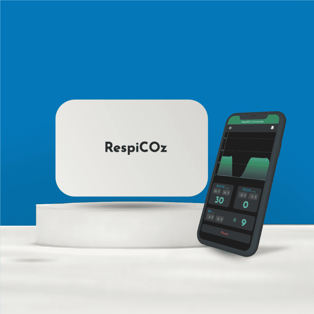
RespiCOz is a portable mainstream capnograph. In an arrest the patient is intubated, so the mainstream sensor sits right at the airway adapter, which is the correct method. The reading appears in seconds with no warm-up, and there is no sampling line to block or water trap to fill during a fast-moving code. It runs on battery, and it pairs with a companion app so the waveform stays in view.
It also stays with the patient after ROSC, through the shift to intensive care or the ambulance, where the airway is still at risk. See EtCO₂ monitoring during transport for that stage.
RespiCOz is a focused respiratory monitor, not a multiparameter monitor, and it is priced in the value middle at ₹60,000 to ₹1,00,000. That makes it realistic to keep one on every resuscitation trolley. For how that compares, see capnograph price in India.
For a current quote for your setting, see the device here.
Frequently asked questions
Why is capnography used in cardiac arrest? It does three things at once. It confirms the airway, it measures the quality of CPR through the blood flow compressions generate, and it signals the return of spontaneous circulation.
What EtCO₂ value shows good CPR? High-quality compressions generally produce an EtCO₂ of at least 10 to 20 mmHg. A value under 10 mmHg is a prompt to improve depth, rate, recoil or to change the compressor.
How does capnography detect ROSC? When circulation returns, more CO₂ reaches the lungs and EtCO₂ rises sharply. A sudden sustained rise is often the first sign of ROSC, before a pulse can be felt.
Can capnography predict survival in cardiac arrest? It carries prognostic information. A low EtCO₂ after 20 minutes of good CPR points to a poor outcome. But guidelines are clear that it must be one part of a multimodal decision, never used in isolation to stop.
What can cause a falsely low EtCO₂ during CPR? Poor compressions, a dislodged or obstructed tube, a disconnected circuit, pulmonary embolism, or the underlying cause of the arrest. Always check these before drawing conclusions from a low value.
Conclusion
Capnography in cardiac arrest gives a resuscitation team the one thing a code usually lacks, an objective, live measure of whether the effort is working.
It confirms the tube. It grades the compressions. It announces ROSC before a pulse. And it offers prognostic information that, handled with care and never alone, helps guide the hardest decisions. Read the trend, act on the number, and respect its limits.
To see how this sits with the other clinical uses of EtCO₂, start with our five capnography use cases.
References
- Part 8: Adult Advanced Cardiovascular Life Support. American Heart Association. Circulation. Waveform capnography for tube confirmation, CPR quality and ROSC. ahajournals.org
- The use of end-tidal carbon dioxide measurement to guide management of cardiac arrest: a systematic review. Resuscitation. EtCO₂ under 10 mmHg after 20 minutes and likelihood of ROSC. sciencedirect.com
- Capnography in Cardiac Arrest. Life in the Fast Lane (LITFL). Prognosis, ROSC detection and confounders. litfl.com




8 thoughts on “Capnography in Cardiac Arrest and CPR”
Pingback: Colorimetric Vs Waveform Capnography: A Safe, Smart Choice
Pingback: 12 Capnography Myths That Still Get Patients Hurt - Brainiac Healthcare - The World's First Smart Capnometer
Pingback: EtCO2 In Pregnancy: The Essential, Safe Normal Range
Pingback: Ambulance Equipment List India: The Complete, Safe Guide
Pingback: Essential Equipment For Emergency Physicians: 5 Safe Buys
Pingback: Capnography Guidelines: A Complete Reference
Pingback: Low EtCO2: Essential Causes And Dangerous Drops
Pingback: Capnography Waveform Chart: A Simple & Clear Field Card