

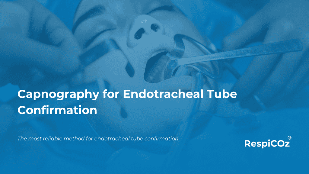
Sustained CO₂ over several breaths confirms the tube is in the airway.
Endotracheal tube confirmation is the moment that decides everything. A tube in the trachea saves a life. The same tube in the oesophagus can end one.
The hard part is that the two can look identical at the bedside. Chest rise, breath sounds and misting in the tube can all appear normal when the tube is in the wrong place. This is why unrecognised oesophageal intubation still causes avoidable death, in the hands of experienced and inexperienced operators alike.
Waveform capnography is the standard for endotracheal tube confirmation. It gives an objective, real-time answer that clinical signs cannot. This guide explains how it confirms placement, what the current guidelines require, and where it can still mislead you.
Key takeaways
- Waveform capnography is the most reliable single method for confirming and monitoring endotracheal tube placement.
- Confirmation depends on sustained exhaled CO₂, not one breath. Look for a repeating waveform over about seven breaths.
- Major guidelines from the AHA and the PUMA airway societies both make it standard practice.
- No sustained trace means the tube is treated as misplaced. The default action is to remove it.
- Capnography can read low or flat in cardiac arrest and low blood flow states. Know the adjuncts.
Why clinical signs are not enough
Intubation is a daily task in theatre, the emergency department and critical care. It is also one of the highest-risk moments in medicine.
Direct visualisation of the tube passing the cords is reassuring. It is not proof. Tubes move during positioning, transfer and transport. Auscultation is subjective and easily misread in a noisy resuscitation.
The 2022 consensus guideline from the Project for Universal Management of Airways states the problem plainly. Preventable oesophageal intubation causes profound hypoxaemia, brain injury and death, and it happens often enough to demand a coordinated response. Clinical judgement alone has been shown, again and again, to be unreliable.
This is the gap that capnography fills.
What capnography actually confirms
Capnography measures carbon dioxide in exhaled breath and displays it as a waveform over time. That waveform is the evidence.
If the tube is in the trachea, each breath returns CO₂ from the lungs. You see a clear, repeating, square-shouldered waveform. If the tube is in the oesophagus, there is little or no CO₂. You see a flat line, or a small trace that fades to zero within a few breaths as any swallowed gas washes out.
The key word is sustained. A single puff of CO₂ can appear from gas in the stomach or pharynx. It disappears fast. A tracheal tube produces CO₂ on every breath and keeps producing it.
For a deeper read on waveform shapes and what they signal, see our guide to capnography waveforms.
What the guidelines say about endotracheal tube confirmation
Two bodies of guidance matter here, and they agree.
Resuscitation. The American Heart Association recommends continuous waveform capnography, alongside clinical assessment, as the most reliable method for confirming and monitoring correct endotracheal tube placement. This is a Class I recommendation, carried through the 2010, 2015, 2020 and 2025 to 2030 ACLS guidelines. In resuscitation it does double duty. It confirms the tube, tracks CPR quality, and flags the return of spontaneous circulation through a sudden rise in EtCO₂.
Anaesthesia and airway management. The 2022 PUMA consensus guideline names the detection of sustained exhaled CO₂ by waveform capnography as the mainstay for excluding oesophageal placement. It is written for every operator, in every setting, not only the operating theatre.
Behind both sits a simple, memorable rule from the Royal College of Anaesthetists 2018 campaign. No trace, wrong place. If you do not see sustained CO₂, you assume the tube is misplaced until proven otherwise.
The seven-breath, two-person check
The PUMA guideline turns the principle into a bedside protocol worth adopting as standard.
- Attach capnography before you commit to the tube.
- Confirm a sustained waveform over about seven breaths.
- Use a two-person verbal check. One person reads the trace aloud, the other confirms.
- If there is no sustained exhaled CO₂, remove the tube.
That last step feels drastic. It is deliberate. The guideline accepts that a few correctly placed tubes will be removed, because the cost of leaving one oesophageal tube in place is catastrophic. The risk-benefit maths favours acting.
When capnography can mislead
An honest guide has to cover the limits. This is where credibility lives.
Capnography needs blood flow to the lungs to carry CO₂ to the alveoli. In cardiac arrest, severe shock or massive pulmonary embolism, pulmonary blood flow drops. A correctly placed tracheal tube can then show a low or even flat reading. This is a false negative, and it has led to correctly placed tubes being pulled.
The practical response is to interpret the trace in context and to keep an adjunct ready.
- Airway ultrasonography. Fast, portable and not dependent on blood flow. It distinguishes tracheal from oesophageal placement in seconds. An Indian study by Roy and colleagues in 2022 compared ultrasound, clinical method and capnography and supported ultrasound as a strong confirmatory tool.
- Colorimetric CO₂ detectors. A chemical indicator changes colour in the presence of CO₂. Useful as a backup, but qualitative only. It cannot show a waveform, cannot monitor over time, and can be fooled by gastric CO₂ for a few breaths.
- Oesophageal detector devices. Helpful in selected cases, with known limits in small children and in obesity.
None of these replaces waveform capnography. They support it when perfusion is the problem.
Colorimetric versus waveform capnography
The two are often confused, so it is worth being precise.
A colorimetric detector answers one question once. Is there CO₂, yes or no. A waveform capnograph answers continuously. It confirms placement, then keeps watching for displacement, disconnection, obstruction and changes in ventilation for as long as the patient is intubated.
For any patient who will be moved, ventilated or monitored beyond the first minute, waveform capnography is the standard. To understand the wider value of continuous EtCO₂ over spot measurements, see our comparison of EtCO₂ and PaCO₂.
Confirmation across settings
The requirement does not change with the room. The equipment often does.
In theatre and the ICU, capnography is built into the anaesthesia machine or the multiparameter monitor. The challenge appears everywhere else. The emergency department, the recovery bay, the general ward, the ambulance and every interhospital transfer. These are exactly the moments when a tube is most likely to move, and often the moments when a full monitor does not travel with the patient.
This is the case for a dedicated, portable capnograph that stays with the patient from intubation through transport. For where a focused device fits against a full monitor, see portable capnograph or multiparameter monitor.
Where RespiCOz fits
RespiCOz is a portable mainstream capnograph built for exactly these moments.
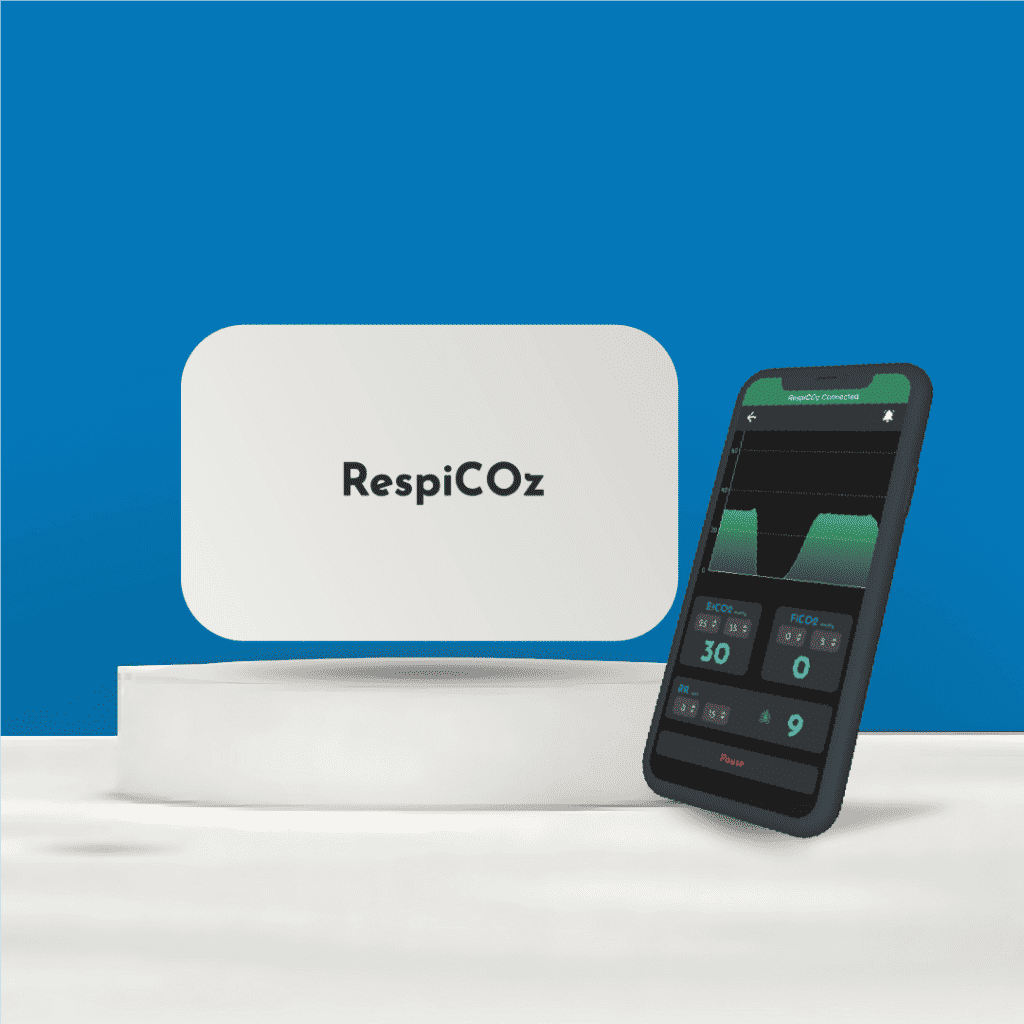
The sensor sits at the airway, so the CO₂ reading is fast and there is no sampling line to block or fill with water. That makes the waveform quick to appear and easy to trust at the point of intubation. It is light enough to stay with the patient into recovery, onto the ward and into the ambulance, so confirmation does not stop when the patient leaves theatre.
It is honest to be clear about what it is. RespiCOz is not a multiparameter monitor. It is a focused respiratory layer that does one job well, wherever the patient is. It is CDSCO-approved, made in India, and it pairs with a companion app for a clear waveform on a screen you already carry.
If that fits a gap in your own practice, you can see the device here.
Frequently asked questions
Is capnography the gold standard for confirming ET tube placement? Yes. Major guidelines describe continuous waveform capnography, used alongside clinical assessment, as the most reliable method for confirming and monitoring correct placement.
How many breaths confirm a tracheal tube? Look for sustained exhaled CO₂ across about seven breaths. A single breath is not enough, because gas from the stomach can produce a brief false trace that fades.
What does a flat capnography line mean after intubation? Treat it as a misplaced tube until proven otherwise. The one important exception is cardiac arrest or very low blood flow, where a correctly placed tube can still read low. Confirm with an adjunct such as airway ultrasound before removing.
Can colorimetric detectors replace waveform capnography? No. They give a one-time yes or no for CO₂. They cannot show a waveform or monitor the tube over time, so they work only as a backup.
Does capnography work outside theatre? Yes, and this is where it matters most. Tubes move during transfer and transport. A portable capnograph lets you confirm and keep monitoring in the ward, recovery and the ambulance.
Conclusion
Endotracheal tube confirmation is not a judgement call. It is a measurement.
Waveform capnography turns a dangerous guess into an objective answer, and then keeps answering for as long as the patient is intubated. Read the trace for sustained CO₂. Apply the seven-breath, two-person check. Know when perfusion can fool you, and keep an adjunct ready.
Do that every time, in every setting, and unrecognised oesophageal intubation stops being a risk you carry.
To see how this sits alongside the other clinical uses of EtCO₂, read our guide to the five capnography use cases that standardise clinical practice.
References
- Panchal AR, et al. Part 8: Adult Advanced Cardiovascular Life Support. American Heart Association Guidelines for CPR and ECC. Circulation. Continuous waveform capnography recommendation (Class I). ahajournals.org
- Chrimes N, Higgs A, Hagberg CA, et al. Preventing unrecognised oesophageal intubation: a consensus guideline from the Project for Universal Management of Airways and international airway societies. Anaesthesia. 2022;77(12):1395–1415. doi:10.1111/anae.15817
- Royal College of Anaesthetists. No trace, wrong place campaign, 2018.
- Roy PS, Joshi N, Garg M, et al. Comparison of ultrasonography, clinical method and capnography for detecting correct endotracheal tube placement. Indian J Anaesth. 2022;66(12):826–831. doi:10.4103/ija.ija_240_22




10 thoughts on “Capnography for Endotracheal Tube Confirmation: 2026 Clinical Guide”
Pingback: Rising Or Falling EtCO2: The Proven Danger Signs
Pingback: Capnography Troubleshooting: Common Errors And How To Fix Them - Brainiac Healthcare - The World's First Smart Capnometer
Pingback: Capnography Troubleshooting: Simple Fixes For Bad Readings
Pingback: Capnography Sensor Placement: A Simple Guide
Pingback: EtCO2 In Pregnancy: The Essential, Safe Normal Range
Pingback: 3 Products Every Anaesthesiologist Must Have - Brainiac Healthcare - The World's First Smart Capnometer
Pingback: Essential Equipment For Emergency Physicians: 5 Safe Buys
Pingback: Ambulance Equipment List: What An ICU On Wheels Needs - Brainiac Healthcare - The World's First Smart Capnometer
Pingback: Ambulance Equipment List India: The Complete, Safe Guide
Pingback: Capnography Waveform Chart: A Simple & Clear Field Card