
Capnography in asthma and COPD is the one situation where the shape of the trace matters more than the number on it.
In obstructive disease, the EtCO2 value can look almost normal while the patient is in serious trouble. But the waveform changes immediately and unmistakably, into the sloping curve clinicians call the shark fin. And when treatment works, that shape recovers before the number does.
This guide explains why the waveform bends, what it tells you about severity, and how to use it to judge whether a bronchodilator is working.
Key takeaways
- Bronchospasm produces the sloping “shark fin” waveform, and the worse the obstruction, the more pronounced it is.
- The shape improves before the number does, so the waveform is the better guide to treatment response.
- A crisp, upright waveform means bronchospasm is not the cause of the distress. Look elsewhere.
- Both a high EtCO2 (above 50 mmHg) and a low one (below 28 mmHg) predict worse outcomes.
- Unlike peak flow, capnography needs no patient effort and works breath by breath.
Why bronchospasm bends the waveform
A normal capnogram is almost a square wave. Bronchospasm destroys that squareness for a specific reason.
In asthma and COPD, the small airways narrow unevenly. Some parts of the lung empty freely, others slowly. So the exhaled breath does not arrive as one clean front. It arrives smeared out in time, with different regions delivering gas with different carbon dioxide contents.
Two things follow. The rise becomes gradual rather than sharp, because dead-space gas and alveolar gas are no longer cleanly separated. And the plateau stops being flat, sloping steadily upward as the slow, CO2-rich regions finally empty.
Together these open up the alpha angle, the corner between the rising limb and the plateau. In severe obstruction that corner disappears altogether, and the trace becomes one continuous sloping curve. That is the shark fin.
For the full catalogue of shapes, see capnography waveforms.
The shark fin as a severity measure
The degree of bend tracks the degree of obstruction. The worse the bronchoconstriction, the steeper the phase III slope, the wider the alpha angle, and the more pronounced the fin.
This has been studied against peak flow and spirometry. The capnogram’s slopes and angles reflect the degree of airway narrowing well, though they are not an exact substitute for spirometry. What they add is something spirometry cannot give: a continuous, effort-independent measurement, breath after breath, in a patient who is too breathless to blow into a peak flow meter.
That last point is the practical one. The sickest asthmatic, the one you most want to measure, is often the one who cannot perform a peak flow.
The insight that matters: shape moves before number
This is the finding most clinicians have not seen, and it changes how you monitor.
When you treat bronchospasm, the waveform’s phase III slope and alpha angle improve significantly, while the EtCO2 value may barely move. One systematic review found the phase III gradient and alpha angle both fell significantly with treatment, while mean EtCO2 was essentially unchanged. A case report tracking an intubated asthmatic through recovery found the phase III slope improved significantly around days three to four, but the EtCO2 value only fell days five to six.
The conclusion is clear. In obstructive disease, the shape is the early indicator, and the number lags behind it.
So if you are watching only the EtCO2 figure, you will be slow to see that your treatment is working, and slow to see it failing.
What the number does tell you
The value is not useless. It carries prognostic weight, in a way that catches people out.
Prehospital data on asthma and COPD exacerbations found that both extremes predict worse outcomes. A high EtCO2, above about 50 mmHg, and a low EtCO2, below about 28 mmHg, were each associated with higher rates of intubation, critical care admission and death.
The logic makes sense. A high value means the patient is tiring and can no longer clear carbon dioxide, which is respiratory failure. A low value means they are hyperventilating hard, which is exhausting and cannot be sustained. A patient who moves from a low value up through the normal range is not necessarily improving. They may be tiring.
That “normalising” EtCO2 in a still-distressed asthmatic is a classic trap. Read it with the waveform and the patient. See high EtCO2 and low EtCO2.
Judging treatment response
This is where capnography earns its keep in obstruction.
Give a bronchodilator such as salbutamol. If it is working, the shark fin straightens back towards a square wave, the alpha angle closes, and the phase III slope flattens. You can watch it happen, in real time, without waiting for a blood gas or a peak flow the patient cannot perform.
If the shape does not improve, the treatment is not working, and you know it early. See drugs that affect EtCO2.
The diagnostic pearl
Here is the one to remember.
If the waveform is crisp and upright, the patient does not have bronchospasm. Whatever is causing their respiratory distress, it is not airway obstruction, and you should look elsewhere.
That single observation redirects a differential fast. A breathless patient with a normal square waveform and a low EtCO2 may have a pulmonary embolism. One with a normal shape and a rising EtCO2 may be tiring from pulmonary oedema. The shape rules obstruction in or out at a glance.
COPD has its own quirks
Two differences are worth knowing.
Chronic CO2 retention. A COPD patient may run a chronically raised baseline EtCO2. Their “normal” is not the standard 35 to 45 mmHg. Judge them against their own baseline and their trend, not the textbook range. See the normal EtCO2 range.
Emphysema. Destruction of the alveolar-capillary bed can distort the plateau further, sometimes producing an unusual downward-trending phase III. It is a structural change, not an acute one.
Where RespiCOz fits
Most published capnography in acute asthma is done on non-intubated patients in the emergency department, sampling through a nasal cannula. That is sidestream or microstream territory, and it is honest to say so.
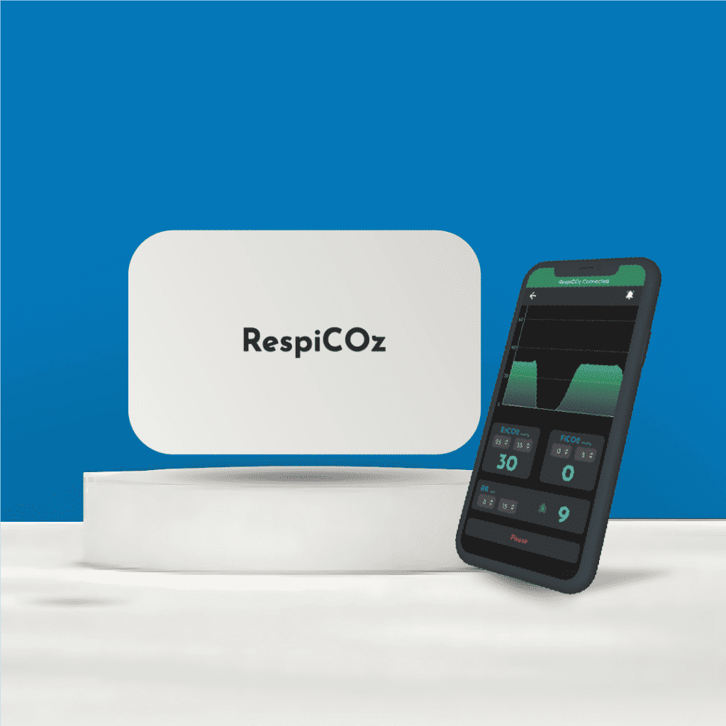
RespiCOz is a mainstream capnograph, so its place in obstructive disease is the intubated and ventilated patient: the severe asthmatic who has required invasive ventilation, and the COPD patient ventilated in intensive care. That is exactly the setting of the case report above, where tracking the phase III slope through recovery revealed improvement days before the number did.
In that setting, RespiCOz gives a fast, direct reading at the airway, with a clear waveform, no sampling line to block with secretions, and FiCO2 to catch rebreathing in the circuit. It is CDSCO-approved, made in India, carries a two-year warranty, and is priced in the value middle at ₹60,000 to ₹1,00,000.
For choosing the right sampling method for your patients, see mainstream vs sidestream capnography. For how RespiCOz compares, see the best handheld EtCO2 monitor guide.
Ready to buy? Request a quote for your hospital here.
Frequently asked questions
What is a shark fin waveform on capnography? It is the sloping capnogram seen in bronchospasm. Uneven narrowing of the small airways smears out the exhaled breath, so the rise becomes gradual and the plateau slopes upward, opening the alpha angle until the trace looks like a shark’s fin.
Does capnography show how severe an asthma attack is? Yes. The steeper the phase III slope and the wider the alpha angle, the worse the obstruction. It reflects airway narrowing well and, unlike peak flow, needs no effort from a breathless patient.
Does EtCO2 rise or fall in an asthma attack? Either. Early on, hyperventilation can lower it. As the patient tires, it rises. Both extremes, above about 50 mmHg and below about 28 mmHg, are associated with worse outcomes, so a “normalising” number in a still-distressed patient may mean exhaustion, not recovery.
How do I know if a bronchodilator is working? Watch the waveform. If the shark fin straightens back towards a square wave and the alpha angle closes, the treatment is working. The shape improves before the EtCO2 number does.
What does a normal, crisp capnography waveform mean in a breathless patient? That bronchospasm is not the cause. A square waveform effectively rules out airway obstruction, so the distress is coming from somewhere else.
Conclusion
Capnography in asthma and COPD works differently from everywhere else, because here the waveform outranks the value.
Bronchospasm bends the trace into a shark fin, and the degree of bend tracks the degree of obstruction. Treatment straightens it, and it straightens before the number falls. A crisp trace means the problem is not obstruction at all. And the value itself carries a warning at both ends, since a tiring patient and a hyperventilating one are both in trouble.
Read the shape first, the number second, and the patient always.
To learn the other waveform shapes, see capnography waveforms.
References
- Capnography: Monitoring Asthma Patients. EMRA. Desynchronisation, parallel heterogeneity and the alpha angle. emra.org
- How to Use End-tidal Capnography to Monitor Asthmatic Patients. ACEP Now. The shark fin waveform in severe attacks. acepnow.com
- Monitoring the Resolution of Acute Exacerbation of Airway Bronchoconstriction in an Asthma Attack Using Capnogram Waveforms. Critical Care Explorations. 2023. Phase III slope improves before EtCO2. journals.lww.com