

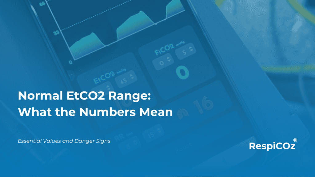
The normal EtCO2 range is 35 to 45 mmHg, which is 4.6 to 6.0 kPa. That single band is the anchor for every capnography reading you will ever take.
Learn it well and the rest becomes simple. A number inside the band means ventilation is on track. A number outside it means something has changed, and the direction tells you where to look. This guide sets out the values, explains why they move, and shows how to read them at the bedside.
Key takeaways
- The normal EtCO2 range is 35 to 45 mmHg (4.6 to 6.0 kPa) in a healthy adult.
- Below 35 mmHg is hypocapnia. Above 45 mmHg is hypercapnia.
- EtCO2 normally reads 2 to 5 mmHg lower than arterial CO2 (PaCO2).
- A low number points to hyperventilation or reduced blood flow to the lungs.
- A high number points to hypoventilation, airway obstruction or rebreathing.
What is a normal EtCO2 range
EtCO2 is the level of carbon dioxide at the end of a breath out. It reflects how well a patient is ventilating.
In a healthy adult at rest, that value settles between 35 and 45 mmHg. This is called normocapnia. It holds across most patients because CO2 production and clearance sit in a steady balance.
Here are the bands you need.
| State | mmHg | kPa | Roughly, vol % |
|---|---|---|---|
| Normal (normocapnia) | 35 to 45 | 4.6 to 6.0 | 4.6 to 6.0 |
| Low (hypocapnia) | below 35 | below 4.6 | below 4.6 |
| High (hypercapnia) | above 45 | above 6.0 | above 6.0 |
The numbers are the same whether you use a sidestream or a mainstream device. For how those devices differ, see our guide to types of capnometers.
Why EtCO2 runs lower than arterial CO2
You will often see EtCO2 sitting just under the arterial value. This is normal.
The difference is the PaCO2 to EtCO2 gradient. In a healthy adult it is about 2 to 5 mmHg. It exists because a small part of each breath fills lung regions that are ventilated but not well perfused, so that gas carries no CO2 and dilutes the sample.
The gradient matters because it widens in disease. Reduced blood flow to the lungs, from pulmonary embolism, shock, blood loss or cardiac arrest, increases the gap. When that happens, EtCO2 understates the true arterial value. This is why EtCO2 is a fast trend tool rather than a direct replacement for a blood gas. For that full comparison, see EtCO2 over PaCO2, and for the dead space behind the gradient, see time and volumetric capnography.
What pushes EtCO2 out of the normal range
A reading outside the band is a clue, not a diagnosis. The value tells you the direction. The clinical picture tells you the cause.
Low EtCO2, below 35 mmHg
- Hyperventilation, where the patient blows off CO2 faster than the body makes it.
- Reduced blood flow to the lungs, from pulmonary embolism, hypovolaemia, hypotension, shock or cardiac arrest.
- A low metabolic state, such as hypothermia.
- Equipment problems, such as a circuit leak, a disconnection or an oesophageal tube, which show a sudden drop or a flat line.
High EtCO2, above 45 mmHg
- Hypoventilation, most often from sedation, opioids or slow recovery from anaesthesia.
- Airway obstruction, such as bronchospasm or COPD, which also changes the waveform shape.
- Increased CO2 production, from fever, sepsis, shivering or malignant hyperthermia.
- Rebreathing of exhaled gas, for example when the soda lime is exhausted. See how that shows up in soda lime and the capnograph.
The normal EtCO2 range in special situations
The 35 to 45 band is the adult resting default. A few common situations shift it, and knowing them prevents false alarms.
- Pregnancy. EtCO2 runs lower, often near 30 mmHg. Progesterone drives a mild chronic hyperventilation, and this is expected.
- Anaesthesia. Ventilation is usually set to hold normocapnia. Over a long case EtCO2 can drift down slowly as metabolism slows.
- CPR. EtCO2 is low during cardiac arrest because blood flow is poor. A value under 10 mmHg suggests chest compressions need to improve. A sudden rise is an early sign of return of spontaneous circulation.
- Children. The target range is broadly the same as adults.
Read the number with the waveform
The value alone is only half the signal. The shape of the trace tells you why.
A normal number with a square waveform is reassuring. A normal number with an abnormal shape, or a changing number over a few breaths, is where the real information sits. Always read the value and the waveform together. Our guide to capnography waveforms covers the common shapes and what each one means.
This is also why a continuous monitor beats a single spot check. A trend shows you the direction of travel. For why that continuous ventilation signal matters, see capnography vs pulse oximetry.
Where RespiCOz fits
Knowing the normal EtCO2 range is only useful if you can see the number where the patient is.
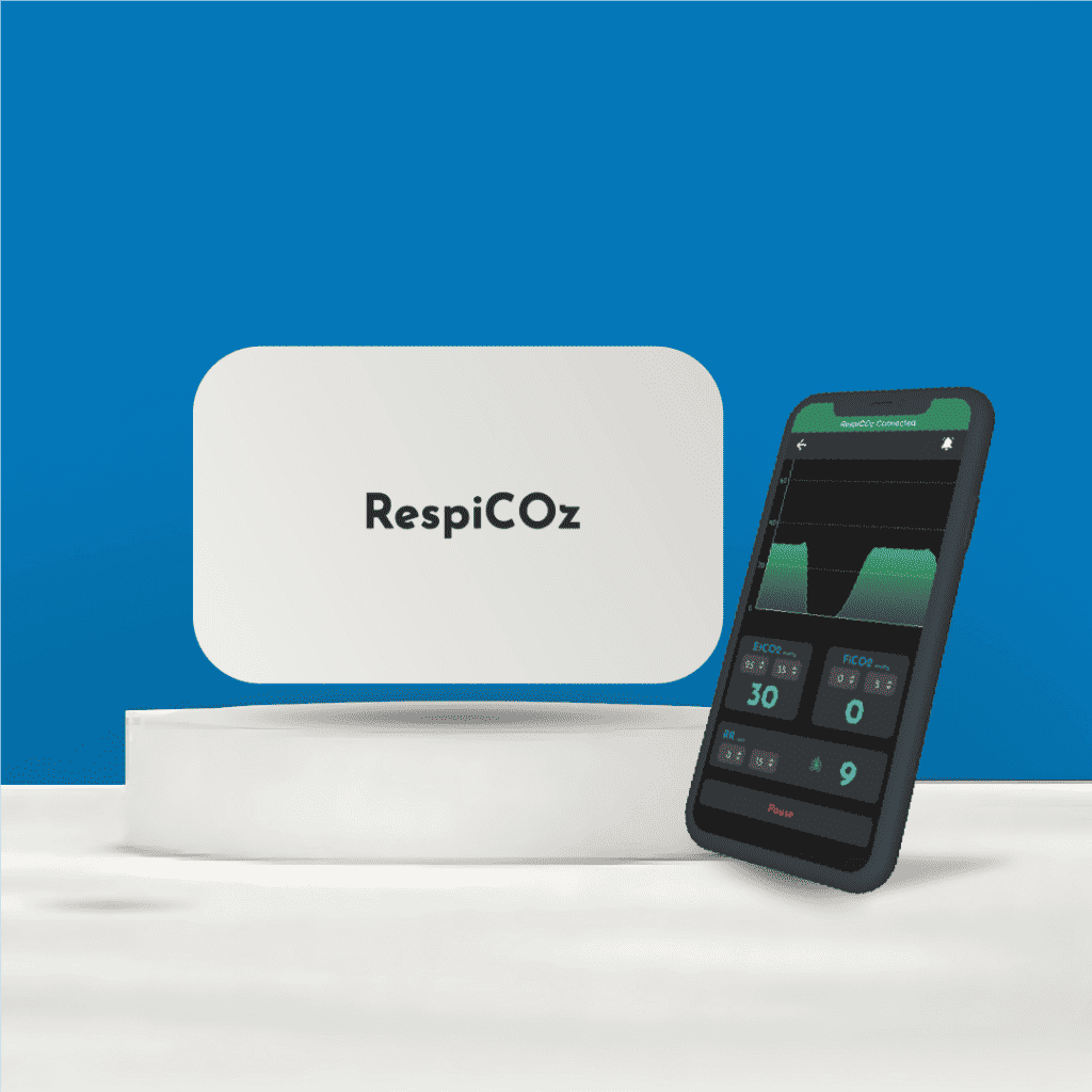
RespiCOz is a portable mainstream capnograph that shows the live EtCO2 value and its trend at the bedside. The sensor sits at the airway, so the reading is fast and there is no sampling line to block or fill with water. It is light enough to stay with the patient through recovery, the ward and transport, which is where a spot reading is often all that is available.
To be clear, RespiCOz is a focused respiratory monitor, not a multiparameter monitor. It does one job well, which is to keep the EtCO2 number and waveform in front of you wherever the patient goes. It is CDSCO-approved, made in India, and pairs with a companion app.
If you want that number at the bedside and not just in theatre, you can see the device here.
Frequently asked questions
What is the normal EtCO2 range? In a healthy adult at rest it is 35 to 45 mmHg, or 4.6 to 6.0 kPa. This is called normocapnia.
Why is EtCO2 lower than arterial CO2? Because of the PaCO2 to EtCO2 gradient, normally 2 to 5 mmHg. A small part of each breath comes from lung regions that are ventilated but not well perfused, which dilutes the CO2 sample.
What does a low EtCO2 mean? A value below 35 mmHg points to hyperventilation or to reduced blood flow to the lungs, such as pulmonary embolism, shock or cardiac arrest.
What does a high EtCO2 mean? A value above 45 mmHg points to hypoventilation, airway obstruction, increased CO2 production or rebreathing of exhaled gas.
What is a normal EtCO2 in pregnancy? It runs lower than the standard range, often around 30 mmHg, because of a mild chronic hyperventilation. This is expected and not a cause for concern on its own.
Conclusion
The normal EtCO2 range, 35 to 45 mmHg, is the anchor every reading is measured against.
Hold that band in your head. A number inside it means ventilation is steady. A number outside it, read with the waveform and the patient, tells you what has changed and where to look next. Low points to hyperventilation or poor blood flow. High points to hypoventilation, obstruction or rebreathing.
Learn the band, watch the trend, and the capnograph stops being a number and becomes a warning system.
To see how this reading is used in practice, start with our five capnography use cases.
References
- End-tidal to arterial CO2 difference. Deranged Physiology. Normal PaCO2-EtCO2 difference of 2 to 5 mmHg. derangedphysiology.com
- The PaCO2-ETCO2 gradient. Don’t Forget The Bubbles. dontforgetthebubbles.com
- Capnography. StatPearls, NCBI Bookshelf. ncbi.nlm.nih.gov





14 thoughts on “Normal EtCO2 Range: What the Numbers Mean”
Pingback: Portable Capnometer Vs Colorimetric: Which Should You Choose? - Brainiac Healthcare - The World's First Smart Capnometer
Pingback: What Does High EtCO2 Mean? - Brainiac Healthcare - The World's First Smart Capnometer
Pingback: High EtCO2: Essential Causes And Danger Signs
Pingback: What Does Low EtCO2 Mean? - Brainiac Healthcare - The World's First Smart Capnometer
Pingback: Low EtCO2: Essential Causes And Dangerous Drops
Pingback: EtCO2 In Trauma: Shock, Head Injury And The Numbers That Matter - Brainiac Healthcare - The World's First Smart Capnometer
Pingback: EtCO2 In Pregnancy: A Different Normal - Brainiac Healthcare - The World's First Smart Capnometer
Pingback: Colorimetric Vs Waveform Capnography: A Safe, Smart Choice
Pingback: Capnography Troubleshooting: Simple Fixes For Bad Readings
Pingback: EtCO2 In Pregnancy: The Essential, Safe Normal Range
Pingback: What Is EtCO2? A Clear, Essential Guide
Pingback: Capnography In Asthma And COPD: The Safe, Smart Guide
Pingback: 5 Types Of Capnometers You Should Know - Brainiac Healthcare - The World's First Smart Capnometer
Pingback: Capnography Waveform Chart: A Simple & Clear Field Card