

Capnography troubleshooting begins with a rule that matters more than any fix in this guide: check the patient before you blame the monitor.
Most capnography problems are simple, and most have a quick remedy. But the one mistake that hurts patients is assuming a real emergency is a technical fault. A flat line is a lost airway until proven otherwise, not a broken sensor. Hold that rule, and everything else here is straightforward.
Key takeaways
- Always rule out a patient problem before you troubleshoot the device.
- A flat trace means a lost airway or a disconnection until proven otherwise.
- Most sidestream faults are a blocked, kinked or loosely connected sampling line.
- Artefacts like the curare cleft and cardiogenic oscillations look alarming but are explainable.
- A mainstream device has no sampling line, so a whole category of faults simply does not arise.
The rule: patient first, device second
Every troubleshooting guide should start here, because the failure mode is predictable.
A monitor shows something alarming. The clinician assumes it is a glitch. Time is lost. Meanwhile the tube is in the oesophagus, or the circuit has come apart, or the patient has stopped breathing.
So before you touch a cable, look at the patient. Is the chest moving. Is the tube where you left it. Is the circuit connected. Only when the patient and the circuit are confirmed do you start treating it as a device problem. Alarm fatigue is real, and it is dangerous, so treat each alarm as true until you have shown otherwise.
Quick reference
| What you see | Likely cause | What to do |
|---|---|---|
| Flat line, no waveform | Disconnection, displaced or blocked tube, oesophageal tube, apnoea | Check the patient and circuit at once. Treat as a lost airway |
| Sudden exponential fall | Collapse in circulation: arrest, embolism, severe hypotension | Clinical emergency, not a device fault |
| Consistently low reading | Leak, over-ventilation, poor perfusion, sample dilution | Check for leaks, check settings, check perfusion |
| Baseline not returning to zero | Rebreathing: exhausted soda lime, failed valve | Change the absorber, check the valves |
| Erratic, inconsistent numbers | Artefact, loose connection, talking patient | Re-seat the connection, look for artefact patterns |
| Notch in the plateau | Curare cleft, muscle relaxant wearing off | Clinical finding, not a fault |
| Ripples at end of expiration | Cardiogenic oscillations | Usually benign. Low PEEP can abolish them |
| Occlusion or blockage alarm | Sampling line blocked with secretions or moisture | Reconnect, then replace the line |
No waveform at all
The most serious presentation, and the one to treat as real.
The causes are a circuit disconnection, a displaced or blocked tube, an oesophageal tube, ventilator failure, or apnoea. All of these are patient emergencies.
Only after you have checked the patient, the tube and every connection should you consider a device cause, such as the sensor not being connected, or the monitor not having zeroed. See endotracheal tube confirmation.
One important exception is worth knowing. In cardiac arrest or very low blood flow, a correctly placed tube can produce a very low or absent trace, because there is almost no circulation to carry CO2 to the lungs. That is a perfusion problem, not a fault.
A low or falling reading
Low readings have both real and technical causes, and you must separate them.
Real causes include hyperventilation, collapsing circulation, pulmonary embolism, and hypothermia. These are covered in what does low EtCO2 mean.
Technical causes include a leak in the circuit or sampling system, dilution of the sample by fresh gas flow, a leak around an uncuffed tube, and sample dilution when a patient is breathing very fast, where a slow-responding device cannot reach the true end-tidal value. That last one matters in children. See capnography response time.
Check for leaks and settings first. But never assume a low reading is technical without confirming perfusion.
A rising baseline
If the waveform does not come back down to zero between breaths, the patient is rebreathing carbon dioxide.
The usual cause in an anaesthesia circuit is exhausted soda lime. A failed one-way valve or an inadequate fresh gas flow will do it too. Change the absorber and check the valves. A monitor that displays FiCO2, the inspired carbon dioxide, shows this directly, because FiCO2 should be near zero. See how soda lime affects the capnograph and what does high EtCO2 mean.
Artefacts that look like problems
Some patterns alarm people unnecessarily. Recognising them saves needless intervention.
Curare cleft. A notch in the top of the expiratory plateau. It appears when a muscle relaxant is wearing off and the patient begins making small spontaneous breaths against the ventilator. It is a clinical finding, not a device fault, and it tells you something useful.
Cardiogenic oscillations. Small ripples at the end of expiration, caused by the beating heart moving nearby lung tissue. They show up at low respiratory rates and are usually physiologically insignificant. But they can cause the monitor to report an inaccurately low EtCO2, and they can even trigger unwanted ventilator breaths, which leads to unnecessary drug doses or circuit fiddling if misread as spontaneous breathing. Applying a low PEEP will often abolish them.
Surgeon interference. Someone leaning on the chest during expiration can produce a dip that mimics a curare cleft.
Chest compression artefact. During CPR, compressions distort the capnogram. This is common, affecting a large share of traces, so read the trend rather than any single breath.
A talking patient, or a loose connection. Both produce erratic, inconsistent readings in a spontaneously breathing patient.
For the full catalogue of shapes, see capnography waveforms.
Sampling line problems (sidestream only)
If you use a sidestream or microstream device, this is where most of your faults will come from.
Occlusion and blockage. Secretions, vomit, blood and simple condensation clog the line. Not every visible droplet needs action, but an occlusion or blockage alarm does. The sequence is: check for kinks and tangles, disconnect and reconnect the line, and if the alarm persists, replace the line.
Loose connections. A sampling line not fully threaded into the monitor causes a distinctive double-humped or “tails-up” waveform, with wrong gas values. This has been misread as low cardiac output, as awareness under anaesthesia, and has led to inhalational anaesthesia being wrongly stopped. Always re-seat the connector before drawing conclusions.
Water traps. They fill, and they must be emptied. A full trap degrades or stops the reading.
Zeroing. Let the device autozero before you attach it to a closed circuit.
Mainstream problems
A mainstream device avoids the whole sampling-line category, but it has its own short list.
Condensation or secretions on the optical window disturb the reading. Keep the adapter clean and positioned so fluid does not pool in it.
Added dead space and weight at the airway. Small in adults, but significant in very small patients.
Positioning. The adapter should sit correctly in the circuit, close to the airway.
For the full trade-off, see mainstream vs sidestream capnography.
The human errors
Not every error is technical. The commonest mistakes are habits.
- Treating the monitor instead of the patient. The number is a clue, not a diagnosis.
- Dismissing alarms. Alarm fatigue kills. Verify, then dismiss.
- Reading the number without the waveform. The shape often names the cause.
- Not establishing a baseline. Attach the monitor and note the normal trace before you need it.
- Assuming EtCO2 equals arterial CO2. In poor perfusion, the gap widens and EtCO2 understates the truth. See the normal EtCO2 range.
How to prevent most problems
- Attach the monitor early and note the baseline waveform.
- Keep connections tight and check them after every patient move.
- For sidestream, keep the line free of kinks and empty the water trap.
- For mainstream, keep the adapter clean and correctly positioned.
- Maintain and calibrate the device according to the manufacturer’s schedule.
- Train the team to read the waveform, not just the number.
Where RespiCOz fits
Look back at the fault list, and one thing stands out. Most everyday capnography faults come from the sampling line.
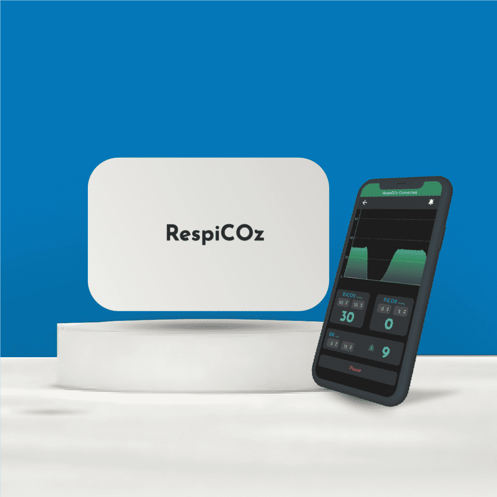
RespiCOz is a mainstream capnograph, so it has no sampling line, no water trap and no Luer connector to work loose. That removes occlusions from secretions, blocked lines, moisture in the tubing, traps that fill up, the double-humped waveform from a loose connection, and the transit delay that comes with drawing a sample. The sensor reads the breath at the airway, so the reading is fast and direct.
It is honest to name what remains. A mainstream adapter must be kept clean of condensation and secretions, and it adds a little dead space, which matters most in very small patients.
RespiCOz shows the value, the waveform and FiCO2 together, which is exactly what you need to tell rebreathing from hypoventilation. It is CDSCO-approved, made in India, with a two-year warranty and a dedicated support team, and priced in the value middle at ₹60,000 to ₹1,00,000. For how it compares, see the best handheld EtCO2 monitor guide.
Ready to buy? Request a quote for your hospital here.
Frequently asked questions
Why is my capnograph not showing a waveform? Treat it as a patient emergency first. The usual causes are a circuit disconnection, a displaced or blocked tube, an oesophageal tube, or apnoea. Only after checking the patient and the circuit should you look for a device fault.
What causes a blocked capnography sampling line? Secretions, vomit, blood and condensation. Check for kinks, disconnect and reconnect the line, and replace it if the occlusion alarm persists. Mainstream devices have no sampling line, so this does not arise.
What is a curare cleft? A notch in the top of the expiratory plateau, seen when a muscle relaxant is wearing off and the patient starts taking small spontaneous breaths. It is a clinical finding, not a device error.
What are cardiogenic oscillations on a capnogram? Small ripples at the end of expiration caused by the beating heart moving lung tissue. They are usually harmless but can cause a falsely low reading or trigger unwanted ventilator breaths. A low PEEP often abolishes them.
Why does my capnograph read low when the patient seems fine? Check for a leak, over-ventilation, or dilution of the sample. But also check perfusion, because poor blood flow lowers EtCO2 for real. Never assume a low reading is only technical.
Conclusion
Capnography troubleshooting is mostly a short list of simple faults: a blocked line, a loose connection, a full water trap, an exhausted absorber, or an artefact that looks worse than it is.
But the discipline that matters is the order you work in. Patient first, circuit second, device third. A flat trace is a lost airway until you prove otherwise, and a low reading may be a failing circulation, not a failing sensor.
Get the order right, learn the handful of artefacts, and the monitor will rarely lie to you.
To read the shapes behind these faults, see our guide to capnography waveforms.
References
- Troubleshooting common capnography challenges. EMS1. Sampling line occlusion, connections and artefact. ems1.com
- Troubleshooting the defect in the sampling line of the sidestream capnograph. NCBI PMC. The double-humped waveform from a loose sampling line. pmc.ncbi.nlm.nih.gov
- Cardiac oscillations in capnograph, abolished with positive end-expiratory pressure. NCBI PMC. pmc.ncbi.nlm.nih.gov




2 thoughts on “Capnography Troubleshooting: Common Errors and How to Fix Them”
Pingback: 12 Capnography Myths That Still Get Patients Hurt - Brainiac Healthcare - The World's First Smart Capnometer
Pingback: Capnography Sensor Placement: A Simple Guide