Carbon dioxide detection in healthcare means measuring CO2 in exhaled breath, primarily through capnography (waveform) and capnometry (numeric EtCO2). Normal adult EtCO2 is 35 to 45 mmHg. The 2025 AHA guidelines recommend continuous waveform capnography as the most reliable way to confirm endotracheal tube placement. This guide covers how the technology works, the major device types, waveform interpretation, troubleshooting, and how medical CO2 detection differs from ambient air quality monitoring.
What Carbon Dioxide Detection Means in Medicine?
Carbon dioxide detection in a clinical setting is the noninvasive measurement of CO2 in a patient’s exhaled breath to assess ventilation. It takes two forms:
Capnometry provides a numeric readout, most often end-tidal CO2 (EtCO2).
Capnography adds a continuous waveform showing CO2 concentration over time (time-based capnography) or over exhaled volume (volumetric capnography).
Most medical CO2 monitors use non-dispersive infrared (NDIR) absorption to measure the gas. Colorimetric detectors offer a simpler, qualitative alternative: they change color when exposed to exhaled CO2 but provide no numbers, trends, or waveforms.
The distinction matters practically. Quantitative waveform capnography is what guidelines endorse for critical decisions like confirming tube placement. Colorimetric devices are backup tools with well-documented limitations.
Watch a quick demo to see how portable capnography works at the bedside.
What You Measure and the Normal Numbers
EtCO2 is the peak carbon dioxide concentration at the end of exhalation. It reflects alveolar CO2 and, indirectly, arterial CO2. In adults with normal lungs, the normal EtCO2 range is 35 to 45 mmHg, corresponding to roughly 4.6 to 5.9% CO2 by volume.
EtCO2 typically runs 2 to 5 mmHg below arterial PaCO2. This gap (the arterial-to-end-tidal gradient) widens when ventilation-perfusion mismatch or dead space increases, as seen in pulmonary embolism, ARDS, or severe COPD. For a closer look at when noninvasive EtCO2 monitoring is preferable to blood gas sampling, read about EtCO2 versus PaCO2.
Another value to watch is FiCO2, the inspired CO2 concentration. It should be near zero. A baseline above roughly 2 mmHg signals rebreathing, meaning the patient is inhaling CO2 that should have been scrubbed or vented from the circuit.
A note on units
Medical carbon dioxide detection uses mmHg or kPa. Ambient and occupational CO2 monitors report in parts per million (ppm). These scales serve entirely different purposes. A normal EtCO2 of 40 mmHg translates to about 52,600 ppm, a number that would sound alarming in an office building but is physiologically normal in exhaled breath.
How CO2 Is Detected in Medical Devices
Most clinical carbon dioxide detection relies on NDIR technology. CO2 absorbs infrared light strongly near a wavelength of 4.26 micrometers. The sensor passes IR light through a gas sample, and a detector measures how much was absorbed. The relationship follows the Beer-Lambert law: higher CO2 concentration means greater absorption and less transmitted light.
NDIR is reliable, specific to CO2, and cost-effective for bedside use. Emerging alternatives like tunable diode laser absorption spectroscopy (TDLAS) and photoacoustic sensors offer faster response and higher sensitivity, but they remain uncommon in routine clinical monitors.
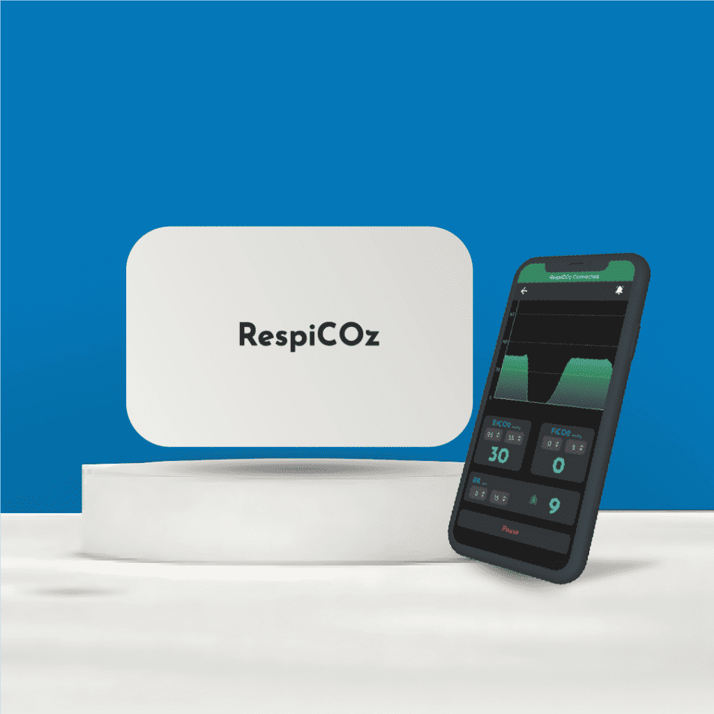
For a practical look at how an NDIR-based capnometer connects to a smartphone in four steps, see the RespiCOz setup walkthrough.
Types of Medical CO2 Detectors
Carbon dioxide detection devices for continuous monitoring come in three main configurations. A fourth option exists for quick qualitative checks.
Mainstream capnography
The IR sensor sits directly at the airway, between the endotracheal tube and the breathing circuit. This delivers near real-time readings with sharp waveforms because gas doesn’t need to travel through tubing.
Tradeoffs: Added weight and dead space at the airway connector. The sensor window can fog from humidity or get contaminated by secretions. Many mainstream sensors require an “adapter zero” when the airway adapter is changed to compensate for optical differences between adapters.
Sidestream capnography
A narrow bore tube continuously aspirates gas from the airway to a remote analyzer. The main advantage is flexibility: sidestream works with non-intubated patients through nasal cannulas, making it popular in sedation suites and emergency departments.
Tradeoffs: Transport delay dampens the waveform and introduces measurement lag. Water traps need regular attention. Line occlusion is a persistent annoyance. Practitioners on paramedic forums consistently cite moisture clogging and sampling line leaks as the top cause of “mystery low EtCO2” or distorted waveforms in the field.
Microstream capnography
A low-flow variant of sidestream, sampling at roughly 50 mL/min instead of the typical 150 to 200 mL/min. Lower flow means less moisture accumulation and reduced dead space impact, making it better suited for neonates and pediatric patients. Even so, users report that microstream sampling hardware can physically interfere with suction or crowd the airway area, so layout around the patient matters.
Colorimetric CO2 detectors
Single-use, battery-free devices that change color (typically purple to yellow) when exposed to exhaled CO2. They offer a quick check for tracheal versus esophageal placement when no waveform device is available.
The limitations are significant. False results occur in low pulmonary blood flow states (like prolonged cardiac arrest), and contamination with gastric acid or epinephrine can cause permanent discoloration that mimics CO2 presence. Colorimetric detectors cannot quantify EtCO2, track trends, or measure respiratory rate.
For a side-by-side comparison with operational checklists, see types of capnometers.
When and Why Clinicians Use CO2 Detection
Confirming and monitoring tube placement
The 2025 AHA Advanced Life Support guidelines recommend continuous waveform capnography as the most reliable method to confirm and monitor endotracheal tube position in adults. A sustained waveform with appropriate EtCO2 values confirms tracheal placement. Loss of the waveform demands immediate action.
Experienced ICU and emergency clinicians treat any rapid desaturation without a convincing capnography waveform as a misplaced tube until proven otherwise. Practitioners on Reddit’s intensive care forums describe waveform capnography as the “real-time truth source” after intubation, trusted above auscultation for immediate confirmation.
Sedation safety
Capnography detects apnea and hypoventilation earlier than pulse oximetry. Oximetry measures oxygen saturation, which can stay normal for minutes after breathing stops, particularly when supplemental oxygen is flowing. Multiple randomized trials show that adding capnography to standard monitoring during procedural sedation reduces hypoxemic episodes. For any setting where patients receive sedation outside the operating room, carbon dioxide detection fills a critical safety gap that oximetry alone cannot cover.
CPR quality and ROSC
During cardiac arrest, EtCO2 reflects the effectiveness of chest compressions by tracking pulmonary blood flow. A sudden sustained rise often signals return of spontaneous circulation (ROSC). Very low persistent EtCO2 has been associated with poor outcomes, though current AHA guidelines caution against using a single cutoff to terminate resuscitation in non-intubated patients.
Reading the Waveform Fast
Time-based capnography produces a characteristic waveform with each breath. Recognizing patterns at the bedside can change outcomes.
Normal waveform
A roughly rectangular shape: baseline near zero, sharp upstroke as exhalation begins, flat alveolar plateau (phase III), a peak at EtCO2, then a rapid downstroke back to baseline during inspiration. This is what you want to see.
Shark fin
A slanted, prolonged upstroke with a poorly defined plateau. This pattern signals obstructive airway disease, typically bronchospasm from asthma or COPD. The airways empty unevenly, so CO2 concentration rises gradually instead of plateauing quickly.
Rising baseline
When the baseline creeps above zero between breaths, the patient is rebreathing exhaled CO2. Common causes include an exhausted soda lime absorber, faulty expiratory valves, or inadequate fresh gas flow.
For more on how absorber chemistry affects the capnograph, read about soda lime and capnography.
Sudden flatline
The waveform drops to zero abruptly. Possible causes include circuit disconnection, extubation, complete airway obstruction, cardiac arrest, or a kinked sampling line. Treat it as a clinical emergency first and a technical issue second. SpO2 may remain normal for minutes after ventilation stops, so waiting on oximetry is dangerous.
For a visual atlas of common and uncommon patterns, explore the capnography waveform guide.
Volumetric Capnography: What It Adds
Standard time-based capnography plots CO2 against time. Volumetric capnography plots CO2 against exhaled volume, and this distinction opens up valuable clinical information. By analyzing the volumetric capnogram, clinicians can partition dead space (using Bohr and Enghoff equations), estimate alveolar dead space fraction, and gain direct insight into V/Q matching.
In ARDS management and ventilator optimization, this granularity helps guide PEEP titration and assess lung recruitment. Volumetric capnography remains underused despite its value, partly because not all devices support it. Learn more about time-based versus volumetric capnography.
Common Pitfalls and How to Fix Them
Carbon dioxide detection technology is reliable when equipment is maintained and operators know what to watch for. These are the problems that come up most often.
Moisture and secretions
The single biggest real-world failure mode. Sidestream lines clog with condensation. Mainstream sensor windows get coated with secretions. Both produce inaccurate readings or erratic waveforms. Fix it by maintaining water traps, replacing wet lines promptly, shielding mainstream adapters from splash during suctioning, and using heated sensors when available. For a deeper explanation, read about moisture-related capnography inaccuracy.
Sampling line leaks and occlusion
In sidestream setups, a kinked or disconnected line produces falsely low or absent EtCO2. If the reading suddenly drops in a patient who appears to be ventilating normally, check the physical line before assuming an airway emergency.
Adapter and sensor zeroing
Many mainstream sensors need an “adapter zero” step whenever the airway adapter is swapped. Skipping it causes baseline drift and misleading values. Make zeroing part of your standard setup checklist.
Neonatal and high-frequency ventilation
In very small patients or during high-frequency oscillatory ventilation, sidestream accuracy degrades because tidal volumes are tiny relative to sampling flow. Know your monitor’s limitations in these populations and consider mainstream alternatives when appropriate.
If your team is standardizing capnography across wards, get a quote on devices built for minimal routine calibration and fast deployment.
Outside the Hospital: Ambient CO2 and Common Detector Confusion
The phrase “carbon dioxide detection” appears in two very different worlds. Mixing them up leads to real misunderstanding.
Ambient and occupational CO2 monitoring
Indoor air quality and workplace safety monitors report CO2 in parts per million. The OSHA 8-hour TWA limit sits at 5,000 ppm, with a short-term exposure limit of 30,000 ppm and an IDLH (immediately dangerous to life or health) threshold around 40,000 ppm. These are environmental safety thresholds. They have nothing to do with clinical EtCO2 targets.
CO is not CO2
Household carbon monoxide alarms detect a completely different gas. Carbon monoxide is a colorless, odorless poison from incomplete combustion. Carbon dioxide is a normal metabolic byproduct. A CO alarm will not detect dangerous CO2 buildup, and a medical capnometer will not detect carbon monoxide poisoning.
The eCO2 problem in consumer sensors
Many inexpensive “CO2” sensors (like the SGP30 or CCS811 chips) do not actually measure CO2. They detect volatile organic compounds and hydrogen, then output an “equivalent CO2” value derived from an algorithm. Engineers and makers consistently warn that these eCO2 readings are unreliable proxies. For accurate carbon dioxide detection in any application, true NDIR sensors are necessary.
Frequently Asked Questions
What is the normal EtCO2 range?
Normal adult EtCO2 falls between 35 and 45 mmHg (roughly 4.6 to 5.9% CO2 by volume). It typically runs 2 to 5 mmHg below arterial PaCO2 in patients with normal lungs.
When should I choose mainstream over sidestream capnography?
Choose mainstream for intubated patients who need the fastest response time and sharpest waveforms. Choose sidestream or microstream for non-intubated patients sampled via nasal cannula, or when you need flexibility across care areas. Expect transit delays with sidestream and plan for regular sampling line maintenance.
Does capnography detect apnea faster than pulse oximetry?
Yes. The capnography waveform flatlines the moment ventilation stops. Pulse oximetry measures oxygen saturation, which can remain normal for several minutes after breathing ceases, especially with supplemental oxygen running. Capnography wins for early detection of ventilatory compromise.
What does a rising baseline mean on the capnogram?
It means the patient is inhaling CO2, a problem called rebreathing. Check the soda lime absorber, expiratory valves, and fresh gas flow. Also verify that the sensor has been properly zeroed if the airway adapter was recently changed.
Can a colorimetric detector replace waveform capnography?
No. Colorimetric detectors serve as a quick adjunct when waveform devices are unavailable, but they cannot quantify EtCO2, trend over time, or detect rate changes. They produce false results during cardiac arrest and when contaminated. Guidelines recommend waveform capnography as the primary tool.
What causes a sudden zero on the capnography waveform?
Possible causes include circuit disconnection, unplanned extubation, complete airway obstruction, cardiac arrest, or a kinked sampling line. Address it as a potential clinical emergency first, then troubleshoot the equipment.
Is carbon dioxide detection the same as carbon monoxide detection?
No. Medical capnometers detect CO2 to assess ventilation. Household CO alarms detect carbon monoxide, a toxic combustion byproduct. The two devices measure entirely different gases and are not interchangeable.
What does eCO2 mean on a consumer air quality sensor?
eCO2 stands for “equivalent CO2.” Many budget sensors estimate CO2 from volatile organic compound readings rather than measuring CO2 directly. These estimates are unreliable for both medical and environmental purposes. For genuine carbon dioxide detection, use true NDIR sensors.
Ready to bring continuous waveform capnography to your clinical team? Explore the 15-day clinical confidence guarantee and trial the technology in your own setting.