

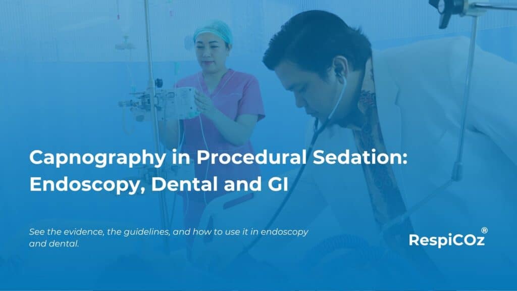
Capnography in procedural sedation is the difference between catching a breathing problem early and finding it late. Sedation makes a patient comfortable. It also slows their breathing, and sometimes stops it.
The most common serious harm during sedation is not cardiac. It is respiratory. A capnograph sees that harm coming, breath by breath, while a pulse oximeter is still showing a reassuring number. This guide covers the evidence, what the guidelines require, and how to use capnography well across endoscopy, dental and GI work.
Key takeaways
- Sedation drugs depress breathing. The commonest serious harm in sedation is respiratory, not cardiac.
- Capnography shows hypoventilation and apnoea within seconds, before pulse oximetry reacts.
- The ASA, ADA and AAOMS now require or recommend capnography for moderate and deep sedation.
- The case is strongest in deep sedation and whenever supplemental oxygen is used.
- It is not perfect. Read the trace with the patient, not on its own.
Why procedural sedation carries respiratory risk
Sedatives and opioids do their job by depressing the central nervous system. That same effect blunts the drive to breathe.
The result can be slow breathing, shallow breathing, airway obstruction or full apnoea. The primary causes of sedation-related harm are drug-induced airway obstruction, aspiration, respiratory depression with hypoventilation, and haemodynamic instability. Most of these are breathing problems.
The danger is that they are quiet. A sedated patient does not complain. Supplemental oxygen keeps the pulse oximeter looking normal for minutes after ventilation has already failed. By the time the number falls, you have lost your early warning. For the full mechanism, see capnography vs pulse oximetry.
What capnography adds during sedation
Capnography measures exhaled carbon dioxide, breath by breath, and shows it as a waveform. It is a direct measure of ventilation.
During sedation this matters in three ways. It flags apnoea the moment breathing stops. It shows hypoventilation as a falling respiratory rate or a changing waveform. And it reveals airway obstruction through the shape of the trace. None of these wait for oxygen to fall first. To read those shapes, see our guide to capnography waveforms, and for the numbers behind them, the normal EtCO2 range.
What the guidelines say about capnography in procedural sedation
The direction of travel is clear, though the societies do not agree on every detail.
Anaesthesia. The American Society of Anesthesiologists standard states that during moderate or deep sedation, the adequacy of ventilation shall be evaluated by observing clinical signs and monitoring for exhaled carbon dioxide, unless precluded by the patient, procedure or equipment. Its 2018 guideline recommends continual capnography to supplement observation and pulse oximetry.
Dentistry and oral surgery. The American Dental Association revised its guidelines so that ventilation for moderate sedation, deep sedation and general anaesthesia must now be assisted by capnography. This was previously required only for intubated patients. The American Association of Oral and Maxillofacial Surgeons has required capnography for moderate and deep sedation in office settings since 2014.
Gastroenterology. This is where the debate sits. The 2018 American Society for Gastrointestinal Endoscopy guideline holds that capnography during moderate sedation has not been shown to improve safety, while still recommending it be considered for deep sedation. So the strongest consensus, across every society, is for deep sedation.
A neutral summary of these positions is in the NCBI evidence brief on capnography for moderate sedation.
What the evidence shows
The guidelines rest on trials, and the trials point one way.
A meta-analysis published in JADA pooled sixteen studies of 3,866 adults in moderate sedation. Adding capnography reduced the risk of hypoxaemia, increased detection of adverse respiratory events, and added no harm.
A separate systematic review in Anesthesia & Analgesia found capnography heralded respiratory depression before oxygen desaturation, especially with supplemental oxygen. Randomised trials in paediatric endoscopy and in adults undergoing ERCP and endoscopic ultrasound showed fewer desaturations, less severe hypoxaemia and fewer apnoea events when capnography was added.
The pattern is consistent. Capnography catches the respiratory event earlier, which buys time to intervene before it becomes hypoxia.
Setting by setting
The principle is the same everywhere. The practical detail changes with the room.
Endoscopy and GI. Deep propofol sedation for colonoscopy, ERCP and endoscopic ultrasound is where the evidence is strongest. Apnoea during a long procedure can go unseen behind supplemental oxygen. Capnography closes that gap.
Dental and oral surgery. Closed-claims data from oral surgery show respiratory distress as the most frequent reason a sedated patient is transferred to an emergency department. Capnography is now a requirement for moderate and deep sedation in these settings, not an optional extra.
Other settings. The same logic applies to bronchoscopy, interventional radiology, cardiology procedures and emergency department sedation. Any sedated, spontaneously breathing patient can slip into respiratory depression without warning.
The limitations to know
An honest guide states the limits. They do not undermine the case. They shape how you use it.
Capnography can mislead. During upper GI endoscopy it can show a false flat line in a minority of patients whose breathing is in fact adequate, which risks unnecessary alarm. Some patients desaturate with a normal capnogram, and some show capnogram changes without desaturation. The lesson is not to distrust the monitor. It is to read the trace alongside pulse oximetry and the patient, never in isolation. Over-reliance on any single alarm leads to alarm fatigue.
How to use capnography well in sedation
Good monitoring is a habit, not a device.
- Attach capnography before the first drug, and note the baseline waveform and rate.
- Watch the trend, not one reading. A falling rate or a changing shape is the early sign.
- Treat any loss of the waveform as apnoea until proven otherwise, and act early.
- Keep supplemental oxygen in mind. It hides desaturation, so the capnogram becomes your main warning.
- Use it with pulse oximetry, clinical observation and someone whose job is to watch the monitor.
Where RespiCOz fits
Most sedation does not happen in a fully monitored operating theatre. It happens in endoscopy suites, dental and oral surgery clinics, GI day-care and procedure rooms. These often have a pulse oximeter, but not built-in capnography.
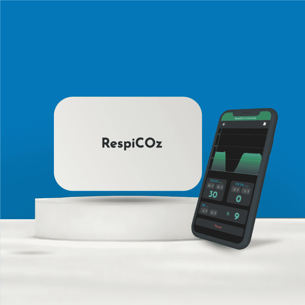
RespiCOz is a portable capnograph that brings the ventilation signal to those rooms. The mainstream sensor sits at the airway, so the reading is fast and there is no sampling line or water trap to replace. It is light enough to stay with the patient from the procedure into recovery and transport, which is where deterioration is often missed. It pairs with a companion app, so the waveform is on a screen you already carry.
It is a focused respiratory monitor, not a multiparameter monitor, and it is priced in the value middle at ₹60,000 to ₹1,00,000. That makes portable capnography realistic for a clinic that cannot justify a full monitor for every room. For how that price compares, see capnograph price in India.
For a current quote for your setting, see the device here.
Frequently asked questions
Is capnography required during procedural sedation? For moderate and deep sedation, the ASA standard calls for monitoring exhaled carbon dioxide, and the ADA and AAOMS require capnography in dental and oral surgery settings. The strongest consensus across all societies is for deep sedation.
Why is capnography better than pulse oximetry during sedation? It detects apnoea and hypoventilation within seconds, before oxygen levels fall. Pulse oximetry is a late warning, especially when the patient is on supplemental oxygen.
Is capnography needed for moderate sedation or only deep sedation? Deep sedation has strong, consistent support across societies. For moderate sedation, the ASA and ADA recommend it, while the gastroenterology guideline is more cautious. Many find the earlier warning worth it either way.
What causes most harm during procedural sedation? Respiratory events. Drug-induced airway obstruction, hypoventilation, apnoea and aspiration are the main causes of sedation-related morbidity, ahead of cardiac problems.
Can capnography give false alarms during sedation? Yes. It can show a false flat line in a minority of patients during upper GI endoscopy. Read it with pulse oximetry and the patient rather than acting on the trace alone.
Conclusion
Capnography in procedural sedation earns its place by seeing the one thing that hurts patients most, a breathing problem, before anything else does.
The evidence shows fewer desaturations and earlier intervention. The guidelines, led by the ASA, ADA and AAOMS, now expect it for moderate and deep sedation. The limits are real, so you read the trace with the patient and the pulse oximeter, not on its own.
Bring capnography into the rooms where sedation actually happens, and the quiet respiratory events stop being quiet.
To see how this sits with the other clinical uses of EtCO2, start with our five capnography use cases.
References
- Evidence Brief: Capnography for Moderate Sedation in Non-Anesthesia Settings. NCBI Bookshelf. Society positions on capnography during moderate sedation. ncbi.nlm.nih.gov
- Benefits and harms of capnography during procedures involving moderate sedation. Journal of the American Dental Association. 2018. Meta-analysis of 16 studies. jada.ada.org
- Continuous Pulse Oximetry and Capnography Monitoring for Postoperative Respiratory Depression and Adverse Events. Anesthesia & Analgesia. 2017. journals.lww.com




3 thoughts on “Capnography in Procedural Sedation: Endoscopy, Dental and GI”
Pingback: Do Small Clinics Really Need Capnography? - Brainiac Healthcare - The World's First Smart Capnometer
Pingback: Capnography For Small Clinics: An Essential Safety Net
Pingback: Drugs That Affect EtCO2: Essential Facts And Danger Signs