Capnography myths survive because they all sound sensible. Each one is a small, plausible shortcut, and each one has led someone to act on a number they should have questioned, or to ignore one they should have acted on.
Here are twelve of them, and what the evidence actually says. Every correction links to a fuller explanation if you want to dig further.
Key takeaways
- A flat line is a lost airway until proven otherwise, not a broken monitor.
- A normal pulse oximeter reading does not mean the patient is breathing.
- EtCO2 is not the same as arterial CO2, and the gap changes with the patient.
- A high EtCO2 is sometimes deliberate. A low one is sometimes an emergency.
- The waveform frequently tells you more than the number does.
Myth 1: “The waveform is flat, so the monitor must be broken”
The truth: it is a lost airway until you prove otherwise.
This is the most dangerous myth on the list. A flat trace means a disconnection, a displaced or blocked tube, an oesophageal tube, or apnoea. Every one of those is a patient emergency.
Check the patient and the circuit first. Only then consider a device fault. Alarm fatigue is real, and it kills. See capnography troubleshooting.
Myth 2: “The oxygen saturation is 98%, so the patient is breathing fine”
The truth: pulse oximetry measures oxygen, not breathing.
Oxygen is stored in a reserve in the lungs and blood. That reserve keeps SpO2 looking normal for minutes after ventilation has failed, and supplemental oxygen makes the delay longer still.
A patient can be apnoeic with a comfortable-looking oximeter. Capnography shows it within seconds. See capnography vs pulse oximetry.
Myth 3: “EtCO2 is basically the same as arterial CO2”
The truth: it normally runs 2 to 5 mmHg lower, and the gap moves.
The gradient exists because of alveolar dead space. And it is not fixed.
It widens in shock, pulmonary embolism, trauma and chest injury, so EtCO2 understates the true arterial value exactly when the patient is sickest. It narrows to almost nothing, and can even reverse, in pregnancy.
So EtCO2 is a fast trend, not a substitute for a blood gas. See EtCO2 over PaCO2 and EtCO2 in pregnancy.
Myth 4: “I saw CO2, so the tube is in”
The truth: one breath proves nothing. You need sustained CO2.
Gas swallowed into the stomach, or carbonated drinks, or vigorous bag-mask ventilation, can all produce a brief puff of CO2 from an oesophageal tube. It fades within a few breaths.
Guidance calls for sustained exhaled CO2 across roughly seven breaths, with a two-person verbal check. A decrementing trace that fades to zero is the classic signature of a tube in the wrong place. See endotracheal tube confirmation.
Myth 5: “A colorimetric detector is basically the same thing”
The truth: it is a pH indicator, and it can be fooled.
A colour-change detector does not measure carbon dioxide directly. It responds to acidity. Which means gastric acid, adrenaline solution, atropine and surfactant can all turn it, falsely suggesting the tube is in the airway.
It also gives no number, no waveform, no trend and no alarms, and moisture disables it within minutes. It is a backup, not a monitor. See colorimetric vs waveform capnography.
Myth 6: “A high EtCO2 is always bad”
The truth: sometimes it is deliberate.
In lung-protective ventilation, particularly in ARDS, low tidal volumes are used to avoid injuring the lungs. The price is a rising carbon dioxide, and it is accepted on purpose. It is called permissive hypercapnia, and a raised CO2 is tolerated as long as the pH holds.
Context decides whether a high number is a problem. See what does high EtCO2 mean.
Myth 7: “A low EtCO2 means the patient is hyperventilating”
The truth: it can mean they are dying.
Carbon dioxide has to be carried by blood to the lungs. If circulation collapses, EtCO2 falls even though ventilation has not changed. Cardiac arrest, massive pulmonary embolism, severe haemorrhage and shock all do this.
The question is always: ventilation, or perfusion? See what does low EtCO2 mean.
Myth 8: “Hyperventilate the head injury to bring the pressure down”
The truth: it increases mortality.
Prehospital data on severe traumatic brain injury found that ventilating to an EtCO2 below 35 mmHg was associated with significantly higher 30-day mortality, and that finding held even in patients with signs of cerebral herniation.
Low CO2 constricts cerebral vessels and starves an already injured brain of blood flow. Target 35 to 45 mmHg. See EtCO2 in trauma.
Myth 9: “EtCO2 under 10 after 20 minutes means stop CPR”
The truth: it is one input, never the decision.
A persistently low EtCO2 after prolonged high-quality CPR does carry a poor prognosis. But guidance is explicit that it may form one part of a multimodal decision and must never be used in isolation.
A low value can also come from poor compressions, a dislodged tube, a disconnected circuit or a pulmonary embolism. During CPR, a value under 10 mmHg is a prompt to improve the compressions, not permission to stop. See capnography in cardiac arrest.
Myth 10: “The number is what matters”
The truth: in obstruction, the shape moves first.
In asthma and COPD, bronchodilator treatment improves the phase III slope and alpha angle significantly while the EtCO2 value barely moves. The waveform recovers days before the number does.
And a crisp, upright waveform effectively rules out bronchospasm, which redirects a differential in one glance. See capnography in asthma and COPD and capnography waveforms.
Myth 11: “Capnographs need regular calibration”
The truth: most modern devices need none.
Modern NDIR capnographs are solid-state and factory-calibrated, with an internal reference channel that corrects for drift as the light source ages. Many manufacturers state that no routine calibration is required.
What they do need is correct zeroing in clean room air, with the adapter fitted, away from the patient and away from your own breath, plus a periodic gas span check to verify accuracy. See capnograph maintenance.
Myth 12: “Capnography is too expensive for a small clinic”
The truth: small settings need it most, and it now costs a fraction of a full monitor.
Office-based and small settings carry a substantially higher risk of adverse events than larger facilities, with less capacity to rescue a deteriorating patient. The monitor matters more where help is furthest away.
And the price objection has collapsed. A multiparameter monitor starts around ₹2,00,000. A portable capnograph runs ₹60,000 to ₹1,00,000 and can cover several rooms. Evidence from sedation monitoring shows capnography reduces acute events and can produce a net saving per procedure. See capnography for small clinics and capnograph cost of ownership.
Two more worth knowing
“Mainstream is more accurate than sidestream.” Not really. Both read EtCO2 accurately and correlate closely. The real differences are patient type, response speed and running cost. See mainstream vs sidestream capnography.
“Capnography can replace lactate in sepsis.” No. It is a good screening tool, with EtCO2 falling as lactate rises, but changes in EtCO2 during treatment do not track changes in lactate. Screen with it. Do not resuscitate to it. See EtCO2 in sepsis.
Where RespiCOz fits
Most of these myths share a root cause: reading a number without the waveform, or having no continuous monitor at all.
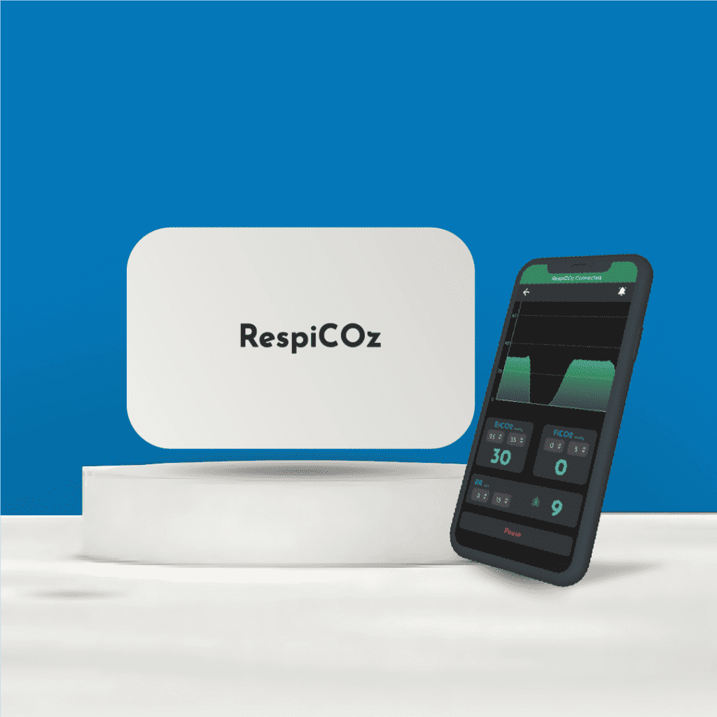
RespiCOz is a portable mainstream capnograph that shows the EtCO2 value, the waveform and the trend together, plus FiCO2 to catch rebreathing directly. It travels with the patient, so monitoring does not stop at the theatre door. As a waveform device it is not fooled by acidity the way a colorimetric detector is, and as a mainstream device it has no sampling line or water trap to fail.
It is CDSCO-approved, made in India, carries a two-year warranty with a dedicated support team, and is priced in the value middle at ₹60,000 to ₹1,00,000. It is a focused monitor for airway-secured patients; free-breathing patients on a nasal cannula need sidestream. For how it compares, see the best handheld EtCO2 monitor guide.
Ready to buy? Request a quote for your hospital here.
Frequently asked questions
Does a flat capnography line mean the monitor is broken? No. Treat it as a lost airway, a disconnection or apnoea until proven otherwise. Check the patient and circuit before you suspect the device.
Is EtCO2 the same as arterial CO2? No. EtCO2 normally reads 2 to 5 mmHg lower. That gap widens in shock, trauma and embolism, and narrows or even reverses in pregnancy, so it cannot be treated as interchangeable.
Is a high EtCO2 always dangerous? No. In lung-protective ventilation for ARDS, a raised carbon dioxide is deliberately tolerated to protect the lungs. It is called permissive hypercapnia.
Should you stop CPR if EtCO2 stays below 10 mmHg? Not on that basis alone. Guidance says a persistently low value may form one part of a multimodal decision but must never be used in isolation, because poor compressions, a dislodged tube or an embolism can all cause it.
Do capnographs need routine calibration? Most modern NDIR devices do not. They need correct zeroing in room air and a periodic gas span check to verify accuracy. Always follow your device’s manual.
Conclusion
Capnography myths persist because each one contains a grain of sense. CO2 usually does mean the tube is in. A low number usually does mean hyperventilation. A high one usually is a problem.
But medicine happens at the edges of “usually”, and that is exactly where these beliefs fail. So hold the harder discipline: read the waveform alongside the number, ask whether a change is ventilation or perfusion, treat a flat line as an emergency, and never let a single value make a decision on its own.
Get those right, and the capnograph becomes what it should be: the fastest honest signal you have.
To start from first principles, see what is EtCO2.
References
- Capnography and CO2 Detectors. Life in the Fast Lane (LITFL). False positives, false negatives and detector limitations. litfl.com
- Capnography. StatPearls, NCBI Bookshelf. Clinical role, gradient and interpretation. ncbi.nlm.nih.gov
- Part 8: Adult Advanced Cardiovascular Life Support. American Heart Association. Circulation. Waveform capnography and the multimodal decision to terminate resuscitation. ahajournals.org