EtCO2 monitoring in non-intubated patients uses a CO2-sampling nasal cannula and a sidestream monitor to watch breathing in patients who are sedated, recovering or receiving opioids. It catches hypoventilation and apnoea earlier than pulse oximetry, especially when the patient is on oxygen.
It is also harder to do well than in intubated patients. Mouth breathing, oxygen flow and loose cannulas all lower the reading. This guide covers why to monitor, where, how to set it up, how to read the values and how to fix common problems.
Key takeaways
- In a meta-analysis, respiratory depression during sedation was 17.6 times more likely to be detected with capnography [1].
- Supplemental oxygen can hide hypoventilation from the pulse oximeter. Capnography does not [2].
- Use a sidestream or microstream monitor with a CO2-sampling nasal cannula, ideally one with an oral scoop [3].
- Expect EtCO2 to read lower than arterial CO2. Watch the trend, the waveform and the breathing rate, not one number [4].
- Guidelines differ: the ASA recommends capnography during moderate sedation, while the ASGE does not require it for endoscopy [5][6].
Why monitor EtCO2 without an airway device?
Sedatives and opioids slow breathing before oxygen levels fall. Capnography sees the slowing directly. A study cited in Respiratory Care found hypoventilation came before oxygen desaturation in about 75% of cases, by an average of 1.7 minutes [3].
Supplemental oxygen makes the pulse oximeter slower to react. In a study of patients made to hypoventilate, desaturation below 90% was four times more common on room air than on supplemental oxygen [2]. The authors concluded that pulse oximetry detects hypoventilation reliably only on room air [2].
The evidence for better detection is strong. Whether it improves outcomes is less settled. A meta-analysis of 13 randomised trials found less oxygen desaturation when capnography was added to standard monitoring during sedation [7]. Other reviews found more respiratory events detected but no clear change in serious adverse events [8]. Read more in capnography vs pulse oximetry and capnography in procedural sedation.
What do the guidelines say?
| Body | Position |
|---|---|
| American Society of Anesthesiologists (2018) | Continually monitor ventilation with capnography during moderate sedation, unless the patient, procedure or equipment prevents it [5] |
| Association of Anaesthetists (2021) | Use capnography during procedural sedation whenever the patient stops responding to voice [9] |
| American Society for Gastrointestinal Endoscopy | Does not regard capnography as essential for endoscopy under moderate sedation [6] |
An Indian Journal of Anaesthesia editorial discussed this disagreement between societies in 2018 [6]. Follow your hospital’s sedation policy, and use capnography wherever sedation may deepen.
Where it is used
- Procedural sedation: emergency department, radiology, cath lab.
- Endoscopy and bronchoscopy suites.
- PACU and recovery. See post-operative capnography.
- Wards with patients on opioids. See opioid-induced respiratory depression.
- Emergency department: asthma and COPD. See capnography in asthma and COPD.
Which equipment do you need?
Use a sidestream or microstream capnograph. Mainstream sensors are mostly used in intubated patients, while sidestream works for both [10]. Microstream draws only 50 ml/min, which reduces moisture in the line and suits small patients [11].
Choose a CO2-sampling nasal cannula made for your monitor. Designs with an oral scoop sample from the mouth as well as the nose, which helps when patients mouth-breathe [3]. Some cannulas also deliver oxygen through separate prongs. See our types of capnometers guide for the differences.
Setup, step by step
- Choose the right size cannula for adult, paediatric or infant patients.
- Connect the sampling line to the monitor and let it warm up and zero, following the maker’s instructions.
- Place the prongs in the nostrils and position the oral scoop in front of the mouth. Loop the tubing over the ears.
- Connect oxygen to the oxygen port if needed. Use the lowest flow that keeps saturation on target.
- Check for a waveform before sedation starts. Ask the patient to breathe normally and note the baseline EtCO2 and respiratory rate.
- Set alarms for no-breath (apnoea), low respiratory rate, and high and low EtCO2, following your unit’s protocol.
- Record the baseline so you can compare against it during the procedure.
Reading EtCO2 in non-intubated patients
Readings from a nasal cannula are usually lower than in intubated patients, for three reasons:
- Dilution by oxygen. Supplemental oxygen mixes with the exhaled breath. In healthy volunteers, continuous oxygen flow gave a wide spread of EtCO2 readings, with a median of 31.45 mmHg and a range of 5.4 to 44.7 mmHg [10].
- Mouth breathing and leaks. Air drawn in around a loose cannula or an open mouth dilutes the sample and gives falsely low values [4].
- Shallow breathing. In patients after abdominal surgery, the gap between EtCO2 and arterial CO2 was smallest with deep breathing and the mouth closed [12].
So read three things together:
- The trend. A steady rise or fall matters more than a single value.
- The waveform. A regular waveform confirms breathing. A flat line means apnoea, obstruction or a displaced cannula.
- The respiratory rate. A falling rate is often the first sign of opioid or sedative effect.
As an example of practical thresholds, one emergency department study defined hypoventilation as EtCO2 above 50 mmHg, a rise of 10 mmHg from baseline, or loss of the waveform [13]. Your unit’s alarm limits should come from its own protocol.
Troubleshooting
| Problem | Likely cause | Fix |
|---|---|---|
| Flat line | Apnoea, airway obstruction, or cannula out of place | Look at the patient first. Open the airway, then reposition the cannula |
| Very low EtCO2 with good breathing | Oxygen dilution, mouth breathing, leak | Reduce oxygen flow if safe, use an oral scoop cannula, check connections |
| Noisy or irregular waveform | Talking, movement, secretions | Reassure the patient, check and clear the line |
| Occlusion alarm | Moisture or secretions in the line | Replace the sampling line. See moisture and capnography accuracy |
| No reading at all | Line not connected, monitor not zeroed | Reconnect, let the monitor zero, check the water trap |
More in our capnography troubleshooting guide.
Where RespiCOz fits, and where it doesn’t
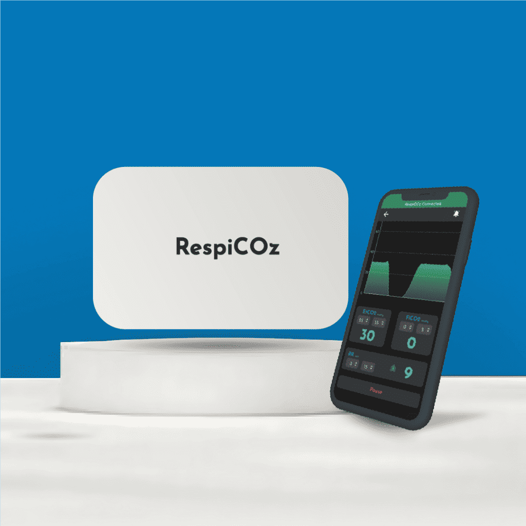
RespiCOz, made by Brainiac Healthcare, the publisher of this guide, is a mainstream capnograph. It is designed for intubated patients, so it is not the right device for nasal cannula monitoring during sedation or endoscopy. For those areas, choose a sidestream or microstream monitor.
If your hospital also needs capnography for intubated patients in the OT, ICU, ambulance or during CPR, RespiCOz shows EtCO2 and the waveform on your phone, at ₹99,990 including GST.
See RespiCOz · Ask us on WhatsApp
Frequently asked questions
Is EtCO2 accurate with a nasal cannula? It tracks trends well but usually reads lower than arterial CO2, especially with oxygen flow or mouth breathing. Use it for trends, breathing rate and waveform presence.
What alarm settings should I use? Follow your unit’s sedation protocol. Typical alarms cover apnoea, low respiratory rate, and high and low EtCO2. Don’t copy limits from one study into practice without local agreement.
Can pulse oximetry alone detect hypoventilation? Only reliably when the patient breathes room air. Supplemental oxygen can delay desaturation and hide hypoventilation [2].
Does oxygen flow affect EtCO2 readings? Yes. Continuous oxygen dilutes the exhaled sample and lowers the reading [10]. Use the lowest flow that keeps saturation on target.
Can a mainstream capnograph monitor non-intubated patients? Mainstream sensors are mostly used in intubated patients [10]. For non-intubated patients, use a sidestream or microstream monitor with a sampling cannula.
Conclusion
EtCO2 monitoring in non-intubated patients gives early warning of hypoventilation and apnoea during sedation, recovery and opioid treatment, often before oxygen saturation falls. Use a sidestream or microstream monitor with a sampling cannula, expect lower readings than in intubated patients, and act on the trend, waveform and breathing rate together.
For intubated patients, see RespiCOz, or request a quote.
References
- Waugh JB, Khodneva YA, Epps CA. Meta-analysis of detection of respiratory events during procedural sedation. Critical Care. 2011;15(Suppl 1):P350.
- Fu ES, et al. Supplemental oxygen impairs detection of hypoventilation by pulse oximetry. Chest. 2004;126(5):1552–8.
- Capnographic oxygen masks vs flow-directed nasal cannula capnography. Respiratory Care. 2017;62(1):78.
- OpenAnesthesia. Capnography.
- American Society of Anesthesiologists. Practice guidelines for moderate procedural sedation and analgesia 2018 (summary).
- Parida S, et al. Standards of care for procedural sedation: focus on differing perceptions among societies. Indian Journal of Anaesthesia. 2018;62(7):493–6.
- Capnography during procedural sedation: meta-analysis of 13 randomised controlled trials. 2017.
- Parker W, et al. Benefits and harms of capnography during procedures involving moderate sedation. Journal of the American Dental Association. 2018;149(1).
- Klein AA, et al. Recommendations for standards of monitoring during anaesthesia and recovery 2021. Anaesthesia. 2021.
- JoVE. Nasal cannula capnography sampling line performance.
- Kodali BS. Microstream technology overview. Capnography.com.
- Deep breathing improves EtCO2 monitoring of an oxygen nasal cannula-based capnometry device. Respiratory Care. 2017;62(1):86–91.
- Sivilotti MLA, et al. Capnometry and pulse oximetry during procedural sedation in adults breathing room air. CJEM. 2010;12(5):397–404.