The essential equipment for emergency physicians is defined by a constraint no other speciality faces quite so sharply. You often have no history, no notes, no diagnosis and no time.
So the devices that matter are the ones that either buy you time or answer a question faster than the alternative. The five below do exactly that, and they are listed in the order a resuscitation actually unfolds: secure the airway, prove it is secure, restart the circulation, get access, then work out what is wrong.
Key takeaways
- First-attempt intubation fails roughly 20 to 30 percent of the time in emergency settings.
- A video laryngoscope improves first-pass success, but a view is not proof.
- A capnograph is the proof, and it keeps proving it as the patient moves.
- A defibrillator with monitor treats the rhythm. It does not tell you the cause.
- Intraosseous access and bedside ultrasound cover the two remaining bottlenecks: access and diagnosis.
1. A video laryngoscope
Its job: secure the airway on the first attempt.
Emergency intubation is not elective intubation. The patient is unstarved, often unstable, frequently in a bad position, and there is no chance to postpone. First-attempt failure in emergency departments and intensive care units runs at roughly 20 to 30 percent, and every extra attempt raises the risk of hypoxaemia, hypotension and cardiac arrest.
The evidence on how to improve that has firmed up. The DEVICE trial, a multicentre randomised trial across 17 emergency departments and intensive care units published in the New England Journal of Medicine in 2023, found that video laryngoscopy produced a higher rate of successful intubation on the first attempt than direct laryngoscopy in critically ill adults.
A camera at the blade tip also means the whole team sees the airway, which matters for supervision and for teaching in a department where juniors intubate.
Its blind spot: it gives you a view, not proof. Seeing a tube pass the cords is reassuring and is not confirmation, because tubes move during securing, positioning and transfer. Which is why the next device exists.
2. A capnograph
Its job: prove the tube is in the trachea, and keep proving it.
This is the device that converts a good view into an objective fact. Waveform capnography measures carbon dioxide in every exhaled breath, and sustained CO2 across several breaths is the definitive confirmation that the tube is in the airway rather than the oesophagus.
The requirement is sustained CO2, not a single puff, because gas swallowed into the stomach can produce a brief false trace that fades within a few breaths. See endotracheal tube confirmation.
In an emergency department it does more than confirm a tube:
In cardiac arrest, it grades CPR quality in real time, since EtCO2 reflects the blood flow your compressions generate, and a sudden sustained rise is often the first sign of return of spontaneous circulation, before a pulse can be felt. See capnography in cardiac arrest.
In trauma, a falling reading flags haemorrhagic shock, often before the blood pressure gives way. See EtCO2 in trauma.
In any deteriorating patient, it detects apnoea and hypoventilation within seconds, while a pulse oximeter on supplemental oxygen can look reassuring for minutes. See capnography vs pulse oximetry.
One warning worth stating. A colorimetric colour-change detector is not a substitute. It responds to acidity rather than to carbon dioxide directly, so gastric acid, adrenaline and atropine can turn it falsely, and it cannot monitor over time. See colorimetric vs waveform capnography.
Its blind spot: it measures ventilation, not oxygenation. And in very low blood flow, a correctly placed tube can read low, so the number must be read in context. See what does low EtCO2 mean.
3. A defibrillator with monitor
Its job: treat the rhythm, now.
For a shockable cardiac arrest, time to defibrillation is the single strongest determinant of survival, and it degrades with every minute of delay. Nothing else in the department substitutes for it.
A modern unit is more than a shock box. It gives continuous ECG, pacing and cardioversion, so it also handles the peri-arrest rhythms, the unstable tachyarrhythmias and bradycardias that arrive before an arrest rather than after it.
Its blind spot: it treats the electrical problem. It tells you almost nothing about why the patient arrested, which is why the last device on this list matters so much in the same resuscitation.
4. An intraosseous access device
Its job: get circulation access when the veins have gone.
Access is the quiet bottleneck of emergency medicine. In arrest, shock, severe dehydration, burns and in small children, peripheral veins collapse or simply cannot be found, and a resuscitation can stall entirely while the team hunts for a line.
An intraosseous device puts a needle into the marrow cavity, usually at the proximal tibia or humeral head, in seconds. The marrow space does not collapse, so it works when veins do not, and fluids, blood and resuscitation drugs can all be given through it. Resuscitation guidance places it as the immediate alternative when intravenous access cannot be obtained promptly.
Its blind spot: it solves delivery, not diagnosis. It buys time. It does not tell you what to give.
5. Point-of-care ultrasound
Its job: answer the diagnostic question at the trolley.
Bedside ultrasound has become the emergency physician’s most versatile tool, because it replaces a journey with an answer. In an undifferentiated hypotensive patient it distinguishes hypovolaemic, cardiogenic, obstructive and distributive shock in minutes, by looking at the heart, inferior vena cava, lungs and abdomen.
It identifies pneumothorax, effusion, consolidation and pulmonary oedema, finds free fluid in trauma, and guides central lines and drains, reducing complications. The international recommendations for point-of-care lung ultrasound reflect a large and growing evidence base for exactly these uses.
Its practical advantage is decisive in an emergency department: it reduces the need for chest X-rays and CT, and it means an unstable patient does not have to be moved to get an answer.
Its blind spot: it is entirely operator-dependent. The device is the cheap part. The training is the investment.
Five devices, five blind spots
| Device | Its job | What it cannot tell you |
|---|---|---|
| Video laryngoscope | Secure the airway first time | Whether the tube is actually in the trachea |
| Capnograph | Confirm the airway and ventilation | Oxygenation, and circulation directly |
| Defibrillator with monitor | Treat the rhythm immediately | Why the patient arrested |
| Intraosseous device | Get access when veins fail | What to give through it |
| Point-of-care ultrasound | Diagnose at the bedside | Anything, without a trained operator |
Read down the last column and the sequence explains itself. Each device hands off to the next.
The gap that shows up in transfers
Emergency medicine has a structural problem the other specialities feel less acutely: the patient never stays put.
A resuscitated patient goes from the resuscitation bay to CT, then to theatre, the catheter lab or intensive care, and sometimes into an ambulance for a transfer to another hospital. Every one of those moves is a moment when a tube can migrate and a circuit can disconnect, and it is usually the moment when monitoring is thinnest.
Guidance is clear that capnography should be continuous for a ventilated patient throughout a transfer, with the display visible to the team. A capnography module built into the resuscitation bay monitor cannot do that, because it stays in the bay. See EtCO2 monitoring during transport.
Where RespiCOz fits
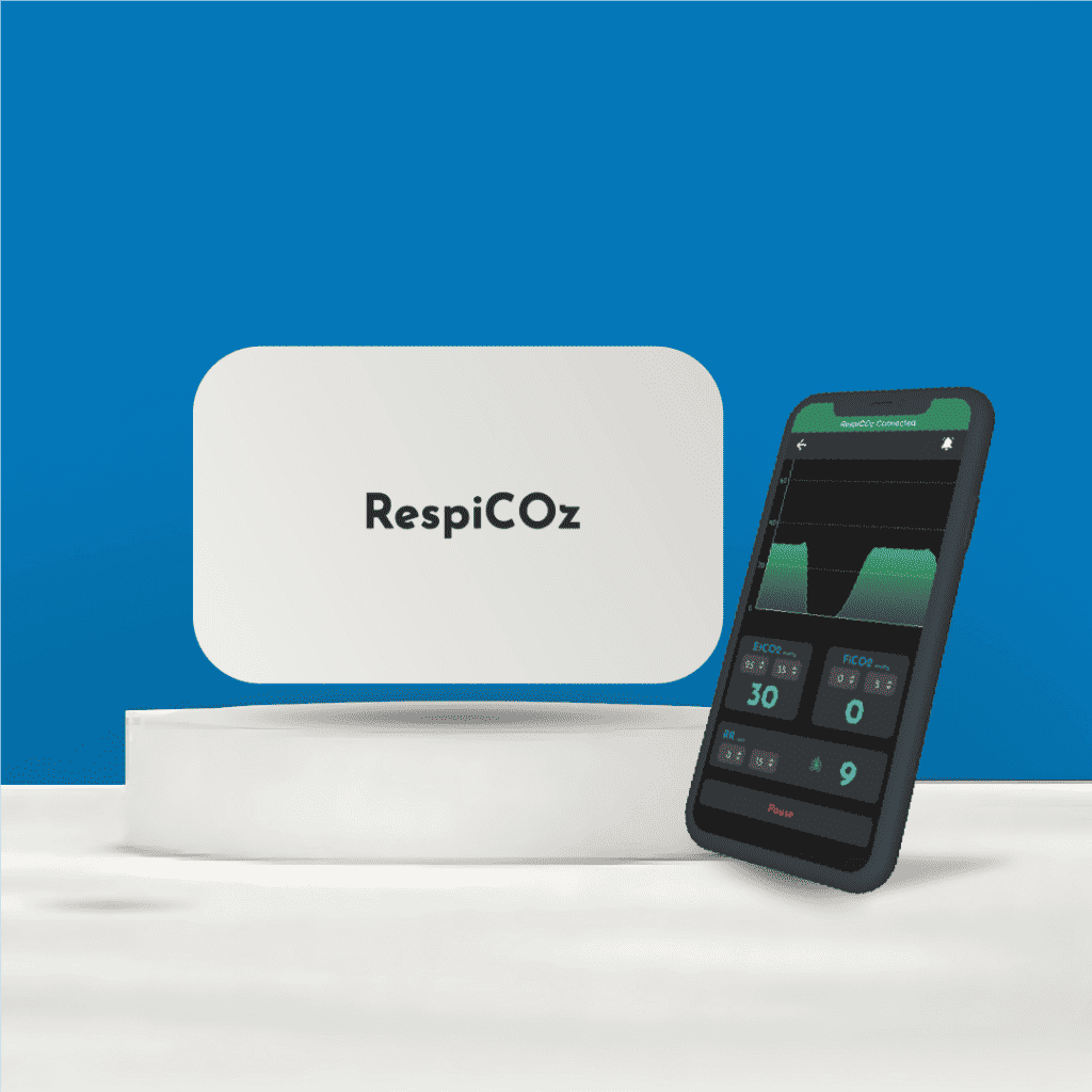
For the capnography slot, RespiCOz is designed for the emergency department reality: intubated patients who move, and no time to fuss with equipment.
The mainstream sensor sits at the airway adapter, so the reading appears in seconds with no warm-up, and there is no sampling line to block with blood, vomit or secretions, which is a genuine problem in emergency airways. There is no water trap to fill in a moving trolley or ambulance either.
It shows the EtCO2 value, the waveform and the trend together, which is what you need during CPR to judge compression quality and to catch the rise that signals return of circulation. It runs on battery and travels with the patient from resus to CT to intensive care, so the airway stays confirmed the whole way.
It is CDSCO-approved, made in India, carries a two-year device warranty with a dedicated technical team, and is priced in the value middle at ₹60,000 to ₹1,00,000. That makes it realistic to keep one on the resuscitation trolley and one for transfers, rather than one for the department. For smaller units weighing the cost, see capnography for small clinics.
To be clear about its scope: RespiCOz is a focused mainstream monitor for airway-secured patients. A spontaneously breathing patient on a nasal cannula, such as a procedural sedation case in the department, needs sidestream or microstream sampling instead.
For how it compares with other portable units, see the best handheld EtCO2 monitor guide.
Ready to buy? Request a quote for your department here.
Frequently asked questions
What equipment does an emergency physician need most? Five cover the first hour: a video laryngoscope to secure the airway, a capnograph to confirm it and monitor ventilation, a defibrillator with monitor for rhythm, an intraosseous device for access when veins fail, and point-of-care ultrasound for bedside diagnosis.
Is a video laryngoscope better than a direct laryngoscope? For critically ill adults, the DEVICE trial found video laryngoscopy produced a higher rate of successful intubation on the first attempt than direct laryngoscopy across emergency departments and intensive care units.
Why do I need a capnograph if I saw the tube pass the cords? Because a view is not proof, and tubes move afterwards during securing, positioning and transfer. Sustained waveform capnography is the objective confirmation, and unlike a visual check it keeps confirming continuously.
Can a colorimetric CO2 detector replace waveform capnography in the ED? No. It responds to acidity rather than carbon dioxide directly, so gastric acid, adrenaline and atropine can produce a false colour change, and it cannot monitor over time or show a waveform.
Why is capnography useful during CPR? It reflects the blood flow your compressions generate, so it grades CPR quality in real time, and a sudden sustained rise is often the earliest sign that circulation has returned, before a pulse is palpable.
Conclusion
The essential equipment for emergency physicians follows the shape of a resuscitation. Secure the airway. Prove it is secure. Treat the rhythm. Get access. Work out what is wrong.
Five devices, and each one hands off to the next, because each is blind exactly where the following one sees. A laryngoscope gives you a view but not proof. A capnograph gives proof but not a diagnosis. A defibrillator treats the rhythm but not the cause.
The one that most often goes missing is the second, not because departments lack capnography, but because it is built into a monitor that stays in the bay while the patient does not. Fix that, and the riskiest minutes of an emergency admission stop being the unwatched ones.
To go deeper on the confirmation itself, see endotracheal tube confirmation.
References
- Prekker ME, Driver BE, Trent SA, et al. Video versus Direct Laryngoscopy for Tracheal Intubation of Critically Ill Adults (DEVICE). New England Journal of Medicine. 2023;389(5):418–429. nejm.org
- Part 8: Adult Advanced Cardiovascular Life Support. American Heart Association. Circulation. Waveform capnography for tube confirmation, CPR quality and ROSC. ahajournals.org
- International evidence-based recommendations for point-of-care lung ultrasound. Intensive Care Medicine. link.springer.com